File:Teacher1925 plate11.jpg
Original file (1,200 × 1,682 pixels, file size: 433 KB, MIME type: image/jpeg)
Plate 11
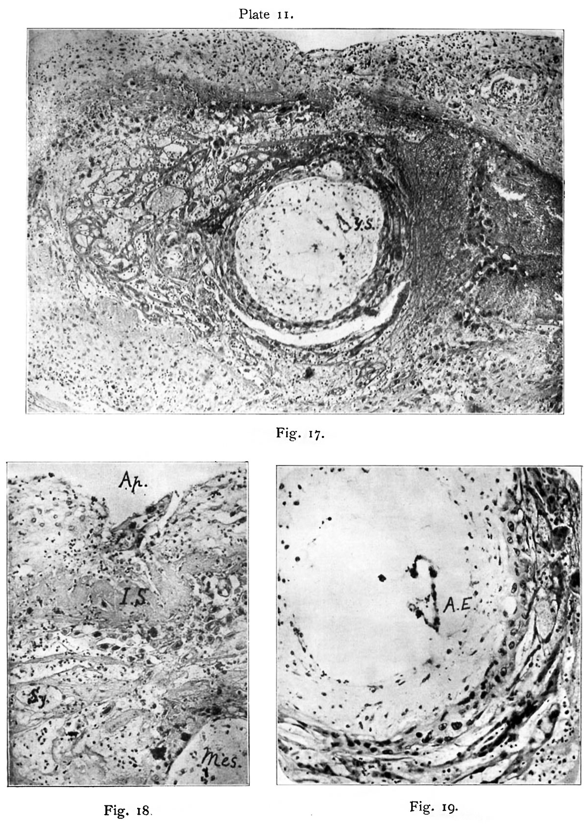
Fig. 17. Teacher—Bryce Ovum No. I.
The whole ovum in Section 4,2,4. X 40. General view showing the decidua capsularis with necrotic zone next the blood in the implantation cavity and the various tissues of the ovum. Compare text fig. 5.
Y.S. The yolk-sac is seen towards one side of the blastocyst imbedded in the mesoderm. Right above it is th-e bulge of the blastocytt wall which is supposed to have been the site of the operculum.
On the left the implantation cavity is occupied by a mass of spun-out spongy syncytium. On the right the spent remains of this are seen flattened against the blastocyst and further out is a line of liberated decidua cells which have been driven towards the middle of the cavity by an inrush of blood. Compare fig. 24, Plate 13. Photograph by J. H. T., 1908.
Fig. 18. The aperture of entrance (Ap.) occupied by remains of the operculum. Section 4.5.4a. Gram-Weigert’s fibrin stain. X 156.
I.S. Internal shield of fibrin with a layer of liberated decidua cells below it. Mes. Mesoderm. Sy. Mass of vacuolated syncytium. Photograph by J. H. T.
Fig. 19. Portion of the blastocyst in Section 4.4.6. X 156 to show.
A.E. The aninio-embryonic sac which is torn. In the photograph masses of deposit from the haemalum complicate the picture. Also shows the mesoderrn, cytotrophoblast and the syncytium (plasmoditrophoblast). Photograph by J. H. T.
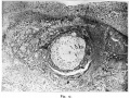
Fig. 17. Teacher—Bryce Ovum No. I.
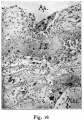
Fig. 18. The aperture of entrance (Ap.) occupied by remains of the operculum
Fig. 19. Portion of the blastocyst in Section 4.4.6.


- Figure Links: text-fig. 1 | text-fig. 2 | text-fig. 3 | text-fig. 4 | text-fig. 5 | plate 1 | plate 2 | plate 3 | plate 4 | plate 11 | fig. 17 | fig. 18 | fig. 19 | Teacher 1925 | Carnegie stage 5 | Carnegie stage 6
| Historic Disclaimer - information about historic embryology pages |
|---|
 |
Reference
Teacher JH. On the implantation of the human ovum and the early development of the trophoblast. (1925) J Obst. Gynaecol. 31(2); 166-217.
Cite this page: Hill, M.A. (2024, April 27) Embryology Teacher1925 plate11.jpg. Retrieved from https://embryology.med.unsw.edu.au/embryology/index.php/File:Teacher1925_plate11.jpg
- © Dr Mark Hill 2024, UNSW Embryology ISBN: 978 0 7334 2609 4 - UNSW CRICOS Provider Code No. 00098GCarnegie Stage 5Week 2
File history
Click on a date/time to view the file as it appeared at that time.
| Date/Time | Thumbnail | Dimensions | User | Comment | |
|---|---|---|---|---|---|
| current | 09:42, 22 November 2016 | | 1,200 × 1,682 (433 KB) | Z8600021 (talk | contribs) | |
| 09:38, 22 November 2016 |  | 1,869 × 2,620 (724 KB) | Z8600021 (talk | contribs) |
You cannot overwrite this file.
File usage
The following page uses this file: