File:Teacher1925 plate06.jpg

Original file (1,874 × 2,510 pixels, file size: 285 KB, MIME type: image/jpeg)
Plate 6
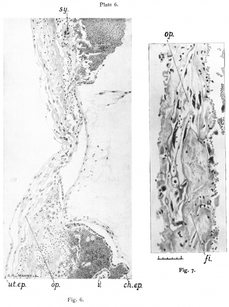
Fig. 6. Teacher-Bryce Ovum No. 2
The operculum in Section 28.2.2. x about 120. It shows the broad, flat and the tearing of the specimen by the air bubble. Ch.ep. Chorionic epithelium.
Sy. Portion of syncytiuni which is also seen in fig. 9, apparently separating the blastocyst from the operculum. V. Villus.
Ut. ep. Remains of uterine epithelium passing in over the surface of the operculum. Compare figs. 8 to 10, photographs. Drawn by A. K.Maxwell.
Fig. 7. Dr. Johnstone’s Ovum No. 1. (Uterine)
The decidua capsularis showing the remains of the operculum (op.) in section 18.2.5. X 320. The blood corpuscles indicate the inner aspect of the clecidua. The operculum is seen as a well defined structure with dark shrivelled nuclei occupying a space in the remains oi‘the decidua. The solid looking mass (fi.) on the inner aspect is part of the internal shield of fibrin. Compare text fig. 2, p. I82, and fig. 11, photograph. Drawn by A. K. Maxwell. Scale: hundredths of mm.
- Figure Links: text-fig. 1 | text-fig. 2 | text-fig. 3 | text-fig. 4 | text-fig. 5 | plate 1 | plate 2 | plate 3 | plate 4 | plate 11 | fig. 17 | fig. 18 | fig. 19 | Teacher 1925 | Carnegie stage 5 | Carnegie stage 6
| Historic Disclaimer - information about historic embryology pages |
|---|
 |
Reference
Teacher JH. On the implantation of the human ovum and the early development of the trophoblast. (1925) J Obst. Gynaecol. 31(2); 166-217.
Cite this page: Hill, M.A. (2024, May 3) Embryology Teacher1925 plate06.jpg. Retrieved from https://embryology.med.unsw.edu.au/embryology/index.php/File:Teacher1925_plate06.jpg
- © Dr Mark Hill 2024, UNSW Embryology ISBN: 978 0 7334 2609 4 - UNSW CRICOS Provider Code No. 00098G
File history
Click on a date/time to view the file as it appeared at that time.
| Date/Time | Thumbnail | Dimensions | User | Comment | |
|---|---|---|---|---|---|
| current | 22:24, 6 June 2018 | | 1,874 × 2,510 (285 KB) | Z8600021 (talk | contribs) | |
| 22:22, 6 June 2018 |  | 2,100 × 2,850 (265 KB) | Z8600021 (talk | contribs) | Plate 6, fig. 6. Teacher-Bryce Ovum No. 2[edit] The operculum in Section 28.2.2. x about 120. It shows the broad, flat and the tearing of the specimen by the air bubble. Ch.ep. Chorionic epithelium. Sy. Portion of syncytiuni which is also seen in fig.... |
You cannot overwrite this file.
File usage
The following page uses this file: