File:T and B lymphocytes EM09.jpg: Difference between revisions
| Line 10: | Line 10: | ||
X 11,500. | X 11,500. | ||
Scale bar 1 micron | Scale bar 1 micron {{Osmium}} | ||
Revision as of 12:41, 23 February 2013
T and B Lymphocyte Electron Micrograph
- Spleen cells labeled to allow a comparison between T and B lymphocytes.
- the undifferentiated lymphocytes appear similar.
Fig. 9 there is a labeled B lymphocyte on the right and an unlabeled, presumably T1, lymphocyte on the left. See T and B Lymphocytes 2 TEM for opposite labeling. Note the resemblance between both cell types (as opposed to the contrast between the two lymphcytes shown in Fig. 7) and the scarcity of organelles in T1 cells.
Labeling in Fig. 9 is done with aMBLA
X 11,500.
Scale bar 1 micron (Stain - Osmium)
- Lymphocyte EM Images: T and B Lymphocytes 1 TEM | T and B Lymphocytes 2 TEM | T Lymphocyte SEM | B lymphocyte 1 TEM | B lymphocyte 2 TEM | B lymphocyte 3 TEM | Plasma Cell TEM | T2 Lymphocyte 1 TEM | T2 Lymphocyte 2 TEM | lymphocyte rosettes | T lymphocyte 1 | T lymphocyte 2 | T lymphocyte 3 | T lymphocyte 4 | T lymphocyte 5 | T lymphocyte 6 | B lymphocyte | B lymphocytes TEM | Immune System Development | Blood
Reference
<pubmed>4563148</pubmed>| PMC2139311
Copyright
Rockefeller University Press - Copyright Policy This article is distributed under the terms of an Attribution–Noncommercial–Share Alike–No Mirror Sites license for the first six months after the publication date (see http://www.jcb.org/misc/terms.shtml). After six months it is available under a Creative Commons License (Attribution–Noncommercial–Share Alike 4.0 Unported license, as described at https://creativecommons.org/licenses/by-nc-sa/4.0/ ). (More? Help:Copyright Tutorial)
File history
Click on a date/time to view the file as it appeared at that time.
| Date/Time | Thumbnail | Dimensions | User | Comment | |
|---|---|---|---|---|---|
| current | 15:11, 22 February 2012 | 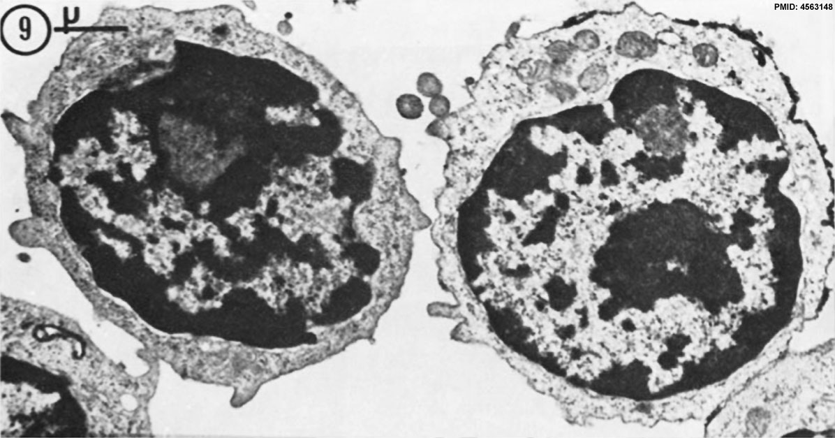 | 1,196 × 627 (137 KB) | Z8600021 (talk | contribs) | T and B_lymphocytes_EM09.jpg |
You cannot overwrite this file.
File usage
The following 9 pages use this file:
- ANAT2241 Lymphatic Tissue and Immune System
- Cardiovascular System - Blood Development
- Immune System - Antibody Development
- Immune System Development
- Lymph Node Development
- SH Lecture - Lymphatic Structure and Organs
- SH Practical - Lymphatic Structure and Organs
- Thymus Development
- Template talk:Lymphocyte images