File:Syngnathidae development 01.jpg
Syngnathidae_development_01.jpg (800 × 325 pixels, file size: 63 KB, MIME type: image/jpeg)
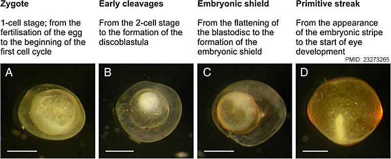
Figure 1 Early embryogenesis
Descriptions of the four stages of the early-embryogenesis period, along with examples for each stage. (A) Animal-pole view of a zygote of N. ophidion ca. 45 min after mating. (B) Animal-pole view of a N. ophidion blastula during early cleavages. (C) Embryonic-shield stage in N. ophidion; the white circle represents the germ ring. (D) Primitive-streak embryo of S. abaster (dechorionated). Scale bars are 0.5 mm.
Reference
<pubmed>23273265</pubmed>| BMC Dev Biol.
Copyright
© Sommer et al.; licensee BioMed Central Ltd. 2012 This article is published under license to BioMed Central Ltd. This is an Open Access article distributed under the terms of the Creative Commons Attribution License (http://creativecommons.org/licenses/by/2.0), which permits unrestricted use, distribution, and reproduction in any medium, provided the original work is properly cited.
File history
Click on a date/time to view the file as it appeared at that time.
| Date/Time | Thumbnail | Dimensions | User | Comment | |
|---|---|---|---|---|---|
| current | 18:06, 19 January 2016 | 800 × 325 (63 KB) | Z8600021 (talk | contribs) |
You cannot overwrite this file.
File usage
The following page uses this file: