File:Sutton1885 fig03.jpg

Original file (1,280 × 1,676 pixels, file size: 258 KB, MIME type: image/jpeg)
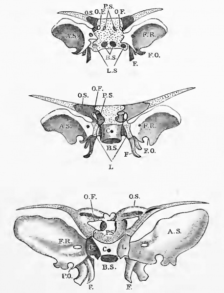
Fig. 3. A series of figures to show the disposition of the various nuclei of the human sphenoid bone
The upper figure shows the relation of the centres to the cartilage; the latter in all cases is represented by dots.
In the middle figure the basisphenoiclal nuclei have coalesced, the orbitosphenoids have joined the presphenoids, and the internal pterygoids have joined the alisphenoids.
In the lowest figure the sphenoid bone is represented as at the eighth month of foetal life.
A.S, Alisphenoid; B.S, basisphenoid ; L , lingulae; F, pterygoids ; O.S , orbitosphenoids ; P.S, presphenoid ; O.F, optic foramen ; F.U, foramen rotundum; F.O, foramen ovale; CC, cranio-pharyngeal canal.
The dorsum sellae at this date is cartilaginous, and therefore it is not represented in the figures. The foramen ovale, until some time after birth, is only a notch in the alisphenoid.
Reference
Sutton JB. On the development and morphology of the human sphenoid bone. (1885) Proc. Zool. Soc. 308: 577-.
Cite this page: Hill, M.A. (2024, April 28) Embryology Sutton1885 fig03.jpg. Retrieved from https://embryology.med.unsw.edu.au/embryology/index.php/File:Sutton1885_fig03.jpg
- © Dr Mark Hill 2024, UNSW Embryology ISBN: 978 0 7334 2609 4 - UNSW CRICOS Provider Code No. 00098G
File history
Click on a date/time to view the file as it appeared at that time.
| Date/Time | Thumbnail | Dimensions | User | Comment | |
|---|---|---|---|---|---|
| current | 12:46, 6 November 2018 | | 1,280 × 1,676 (258 KB) | Z8600021 (talk | contribs) | |
| 12:41, 6 November 2018 | 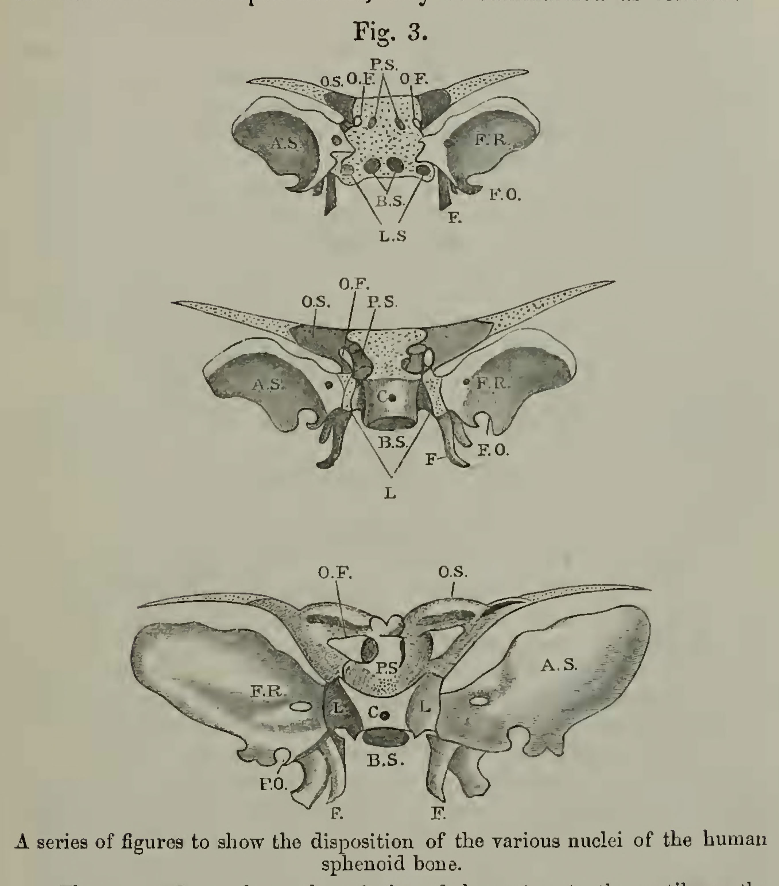 | 2,714 × 3,086 (469 KB) | Z8600021 (talk | contribs) | {{Ref-Sutton1885}} |
You cannot overwrite this file.
File usage
The following page uses this file: