File:Sutton1885 fig01.jpg
From Embryology
Size of this preview: 463 × 599 pixels. Other resolution: 1,000 × 1,294 pixels.
Original file (1,000 × 1,294 pixels, file size: 159 KB, MIME type: image/jpeg)
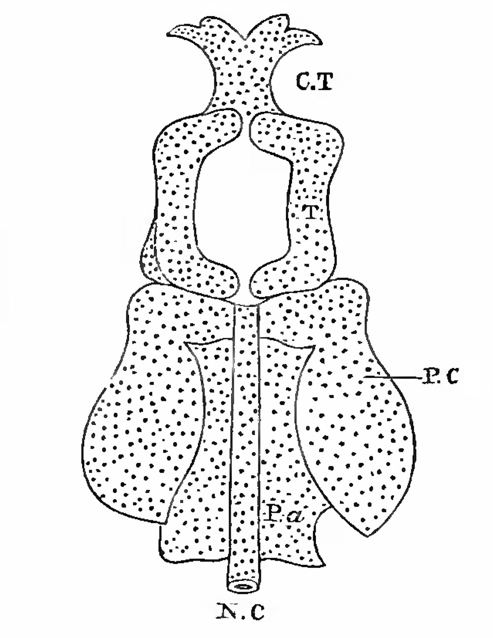
Fig. 1. A diagram to represent the disposition of parts in tlie base of the primitive skull
N.C, Notochord ; Pa, parachordals ; PC, periotic capsules; T, trabeculae; C.T, ethmo-vomerinc region.
Sutton JB. On the development and morphology of the human sphenoid bone. (1885) Proc. Zool. Soc. 308: 577-.
File history
Click on a date/time to view the file as it appeared at that time.
| Date/Time | Thumbnail | Dimensions | User | Comment | |
|---|---|---|---|---|---|
| current | 12:45, 6 November 2018 | | 1,000 × 1,294 (159 KB) | Z8600021 (talk | contribs) | |
| 12:41, 6 November 2018 |  | 2,760 × 2,326 (386 KB) | Z8600021 (talk | contribs) | {{Ref-Sutton1885}} |
You cannot overwrite this file.
File usage
The following page uses this file: