File:Suprascrotal testis.jpg

Original file (1,000 × 751 pixels, file size: 126 KB, MIME type: image/jpeg)
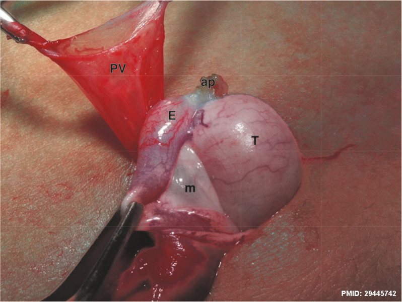
Suprascrotal Testis
A 4-year-old patient with suprascrotal testis presented anatomical relationship between the testis and the epididymis Type I, epididymis attached to the testis at the head and tail.
ap - Testicular appendix
PV - patency of the processus vaginalis
m = mesorchium
T = testis
E = epididymis
- Links: cryptorchidism
Reference
Favorito LA, Riberio Julio-Junior H & Sampaio FJ. (2017). Relationship between Undescended Testis Position and Prevalence of Testicular Appendices, Epididymal Anomalies, and Patency of Processus Vaginalis. Biomed Res Int , 2017, 5926370. PMID: 29445742 DOI.
Copyright
© 2017 Luciano A. Favorito et al. This is an open access article distributed under the Creative Commons Attribution License, which permits unrestricted use, distribution, and reproduction in any medium, provided the original work is properly cited.
Figure 2
Cite this page: Hill, M.A. (2024, April 27) Embryology Suprascrotal testis.jpg. Retrieved from https://embryology.med.unsw.edu.au/embryology/index.php/File:Suprascrotal_testis.jpg
- © Dr Mark Hill 2024, UNSW Embryology ISBN: 978 0 7334 2609 4 - UNSW CRICOS Provider Code No. 00098G
File history
Click on a date/time to view the file as it appeared at that time.
| Date/Time | Thumbnail | Dimensions | User | Comment | |
|---|---|---|---|---|---|
| current | 10:46, 14 October 2018 | | 1,000 × 751 (126 KB) | Z8600021 (talk | contribs) | |
| 10:45, 14 October 2018 | 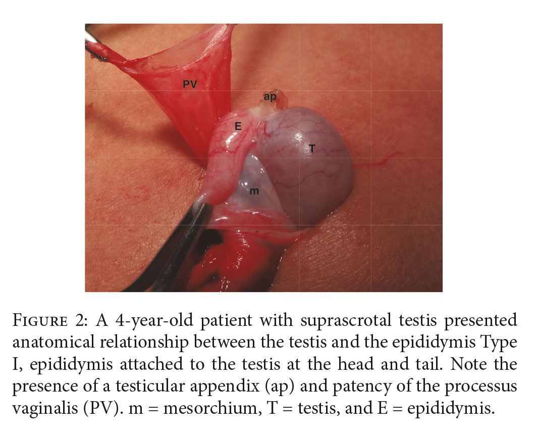 | 1,064 × 857 (138 KB) | Z8600021 (talk | contribs) | ==Suprascrotal Testis== A 4-year-old patient with suprascrotal testis presented anatomical relationship between the testis and the epididymis Type I, epididymis attached to the testis at the head and tail. Note the presence of a testicular appendix... |
You cannot overwrite this file.
File usage
The following page uses this file: