File:Stria Vascularis diagram 2.jpeg
Stria_Vascularis_diagram_2.jpeg (291 × 336 pixels, file size: 46 KB, MIME type: image/jpeg)
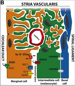
Diagram of the Stria Vascularis, containing the marginal cells, intermediate melanocytes and basal cells. It shows marginal cell extensions intercalating with melanocytes, and the K+ ion channels involved in generating the EP.
Copyright
Copyright © 2015 The Authors Developmental Neurobiology Published by Wiley Periodicals, Inc. This is an open access article under the terms of the Creative Commons Attribution‐NonCommercial License, which permits use, distribution and reproduction in any medium, provided the original work is properly cited and is not used for commercial purposes.
Reference
- Note - This image was originally uploaded as part of an undergraduate science student 2018 project and may contain inaccuracies in either description or acknowledgements. Students have been advised in writing concerning the reuse of content and may accidentally have misunderstood the original terms of use. If image reuse on this non-commercial educational site infringes your existing copyright, please contact the site editor for immediate removal.
File history
Click on a date/time to view the file as it appeared at that time.
| Date/Time | Thumbnail | Dimensions | User | Comment | |
|---|---|---|---|---|---|
| current | 17:57, 4 September 2018 | | 291 × 336 (46 KB) | Z5229132 (talk | contribs) | Diagram of the Stria Vascularis, containing the marginal cells, intermediate melanocytes and basal cells. It shows marginal cell extensions intercalating with melanocytes, and the K+ ion channels involved in generating the EP. ====Copyright==== Copyri... |
You cannot overwrite this file.
File usage
The following 2 pages use this file: