File:Streeter1957 plate04.jpg: Difference between revisions
mNo edit summary |
mNo edit summary |
||
| (One intermediate revision by the same user not shown) | |||
| Line 3: | Line 3: | ||
A renal vesicle will soon form in that part of the metanephric tissue (fig. 26) which lies beneath the ampullae end of a collecting tubule. Successive steps in the development of the tubules are shown in specimens of the several age groups. | A renal vesicle will soon form in that part of the metanephric tissue (fig. 26) which lies beneath the ampullae end of a collecting tubule. Successive steps in the development of the tubules are shown in specimens of the several age groups. | ||
Fig. 26. No. | Fig. 26. No. {{CE576}} | ||
Fig. 27. No. | Fig. 27. No. {{CE8092}} | ||
Fig. 28. No. | Fig. 28. No. {{CE8157}} | ||
Fig. 29. No. | Fig. 29. No. {{CE5537}} | ||
Fig. 30. No. | Fig. 30. No. {{CE6531}} | ||
Fig. 31. No. | Fig. 31. No. {{CE7392}} | ||
Fig. 32. No. | Fig. 32. No. {{CE4638}} | ||
Fig. 33. No. | Fig. 33. No. {{CE8394}} | ||
Fig. 34. No. | Fig. 34. No. {{CE4570}} | ||
{{Streeter1957 figures}} | {{Streeter1957 figures}} | ||
[[Category:Week 8]][[Category:Renal]] | [[Category:Week 8]][[Category:Renal]] | ||
[[Category:Carnegie Embryo 576]][[Category:Carnegie Embryo 8092]] | |||
[[Category:Carnegie Embryo 8157]][[Category:Carnegie Embryo 5537]] | |||
[[Category:Carnegie Embryo 6531]][[Category:Carnegie Embryo 7392]] | |||
[[Category:Carnegie Embryo 4638]][[Category:Carnegie Embryo 8394]] | |||
[[Category:Carnegie Embryo 4570]][ | |||
Latest revision as of 22:07, 30 March 2017
Plate 4. Photographs of sections illustrating uriniferous tubules in the embryos of horizons xix to xxiii
A renal vesicle will soon form in that part of the metanephric tissue (fig. 26) which lies beneath the ampullae end of a collecting tubule. Successive steps in the development of the tubules are shown in specimens of the several age groups.
Fig. 26. No. 576
Fig. 27. No. 8092
Fig. 28. No. 8157
Fig. 29. No. 5537
Fig. 30. No. 6531
Fig. 31. No. 7392
Fig. 32. No. 4638
Fig. 33. No. 8394
Fig. 34. No. 4570
| Historic Disclaimer - information about historic embryology pages |
|---|
 |
- Links: 1 Graph Embryos 11-23 | 4 Eye and optic nerve 19-23 | Plate 1 - Cornea | Plate 2 - Hypophysis
Reference
Streeter GL. Developmental Horizons In Human Embryos Description Or Age Groups XIX, XX, XXI, XXII, And XXIII, Being The Fifth Issue Of A Survey Of The Carnegie Collection. (1957) Carnegie Instn. Wash. Publ. 611, Contrib. Embryol., 36: 167-196.
Cite this page: Hill, M.A. (2024, May 26) Embryology Streeter1957 plate04.jpg. Retrieved from https://embryology.med.unsw.edu.au/embryology/index.php/File:Streeter1957_plate04.jpg
- © Dr Mark Hill 2024, UNSW Embryology ISBN: 978 0 7334 2609 4 - UNSW CRICOS Provider Code No. 00098G[
File history
Click on a date/time to view the file as it appeared at that time.
| Date/Time | Thumbnail | Dimensions | User | Comment | |
|---|---|---|---|---|---|
| current | 17:01, 30 October 2016 | 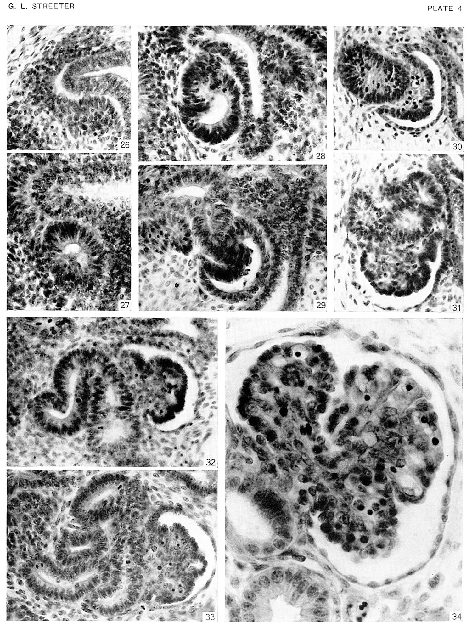 | 1,500 × 1,986 (736 KB) | Z8600021 (talk | contribs) |
You cannot overwrite this file.
File usage
The following page uses this file: