File:Streeter1920 Plate 1.jpg

Original file (758 × 1,000 pixels, file size: 108 KB, MIME type: image/jpeg)
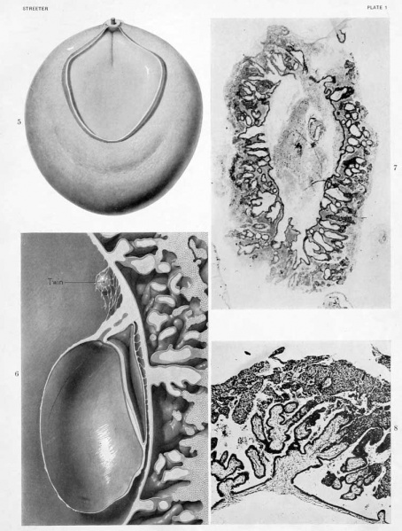
Plate 1 Presomite Embryo
Fig. 5. Frontal view of wax-plate reconstruction of Mateer embryo, showing yolk-sac and embryonic plate outlined by the cut edge of the amnion, which is shown as removed. The stump of the body-stalk can be seen with the allantois at its center. Extending forward from the caudal end of the embryonic plate is a well-defined primitive groove. Enlarged 60 diameters.
Fig. 6. Median view of wax-plate reconstruction of Mateer embryo showing the form of the amniotic cavity and its relation to the chorionic membrane and the yolk-sac. The allantois projects through the body-stalk and is interrupted near its center. In the loose tissue caudal to the body-stalk is an epithelial mass constituting the amniotic vesicle of the twin embryo. Enlarged &) diameters.
Fig. 7. Photograph of section through embryo No. 763, Carnegie Collection. This specimen would belong among those in our group 1 having branched villi. The embryo, consisting of two vesicles, can be seen in the center. The series being incomplete, the definite form of the embryo can not be made out. This specimen is distinctly younger than the Mateer specimen and its viUi are to be compared with those in figure 8. Enlarged 30 diameters.
Fig. 8. Photograph of a typical larger villus of the Mateer specimen, showing transition into trophoblast. For a detail of these structures see text-figure 4. Enlarged 50 diameters.
| Historic Disclaimer - information about historic embryology pages |
|---|
| Pages where the terms "Historic" (textbooks, papers, people, recommendations) appear on this site, and sections within pages where this disclaimer appears, indicate that the content and scientific understanding are specific to the time of publication. This means that while some scientific descriptions are still accurate, the terminology and interpretation of the developmental mechanisms reflect the understanding at the time of original publication and those of the preceding periods, these terms, interpretations and recommendations may not reflect our current scientific understanding. (More? Embryology History | Historic Embryology Papers) |
- Paper Links: Fig 1 | Fig 2 | Fig 3 | Fig 4 | Fig 5 | Fig 6 | Fig 7 | Fig 8 | Fig 9 | Fig 10 | Fig 11 | Fig 12 | Fig 13 | Fig 15 | Fig 16 | Table 1 | Chart 1 | Chart 2 | Chart 3 | Plate 1 | Plate 2 | Plate 3 | Plate 4 | Plate 5 | Plate 6 | Plate 7 | Paper | Contributions to Embryology
Reference
Streeter GL. A human embryo (Mateer) of the pre-somite period. (1920) Contrib. Embryol., Carnegie Inst. Wash. Publ. 272, 9: 389-424.
Cite this page: Hill, M.A. (2024, April 27) Embryology Streeter1920 Plate 1.jpg. Retrieved from https://embryology.med.unsw.edu.au/embryology/index.php/File:Streeter1920_Plate_1.jpg
- © Dr Mark Hill 2024, UNSW Embryology ISBN: 978 0 7334 2609 4 - UNSW CRICOS Provider Code No. 00098G
File history
Click on a date/time to view the file as it appeared at that time.
| Date/Time | Thumbnail | Dimensions | User | Comment | |
|---|---|---|---|---|---|
| current | 09:49, 7 April 2012 |  | 758 × 1,000 (108 KB) | Z8600021 (talk | contribs) | {{Streeter1920a}} |
You cannot overwrite this file.
File usage
The following 3 pages use this file:
