File:Streeter1920 12.jpg
Original file (1,000 × 714 pixels, file size: 48 KB, MIME type: image/jpeg)
| Historic Disclaimer - information about historic embryology pages |
|---|
 |
- Paper Links: Fig 1 | Fig 2 | Fig 3 | Fig 4 | Fig 5 | Fig 6 | Fig 7 | Fig 8 | Fig 9 | Fig 10 | Fig 11 | Fig 12 | Fig 13 | Fig 15 | Fig 16 | Table 1 | Chart 1 | Chart 2 | Chart 3 | Plate 1 | Plate 2 | Plate 3 | Plate 4 | Plate 5 | Plate 6 | Plate 7 | Paper | Contributions to Embryology
Reference
Streeter GL. A human embryo (Mateer) of the pre-somite period. (1920) Contrib. Embryol., Carnegie Inst. Wash. Publ. 272, 9: 389-424.
Cite this page: Hill, M.A. (2024, April 27) Embryology Streeter1920 12.jpg. Retrieved from https://embryology.med.unsw.edu.au/embryology/index.php/File:Streeter1920_12.jpg
- © Dr Mark Hill 2024, UNSW Embryology ISBN: 978 0 7334 2609 4 - UNSW CRICOS Provider Code No. 00098G
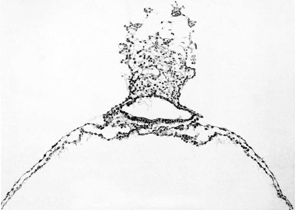
The primitive groove can be seen very clearly in the sections (figs. 12, 13, and 14, plates 2 and 3), and can be traced into the most caudal sections. Where one would expect to find the primitive node the sections become very oblique, so that it can not be outhned with great certainty. It is probably represented, however, in figure 14, plate 3. In this region the ectoderm fuses more or less c; mpletely with the endoderm, and lateral to the point of fusion can be seen a flattened area of mesoderm (fig. 13, plate 3). This mesodermal tissue is in the form of a reticula syncytium, which in most regions is closely attached to the ventral surface of the embryonic plate. It is somewhat more loosely attached to the endoderm by irregular trabeculae. In its more lateral areas it is slightly more condensed into a tissue from which are derived the somites and the more laterally situated unsegmented mesoderm. As to the presence of a head process there seems to be no evidence, although the oblique direction of the sections makes it impossible to rule it out with certaintj'. The most careful scrutiny, however, fails to reveal any sign of a head-process canal.
File history
Click on a date/time to view the file as it appeared at that time.
| Date/Time | Thumbnail | Dimensions | User | Comment | |
|---|---|---|---|---|---|
| current | 09:43, 7 April 2012 | | 1,000 × 714 (48 KB) | Z8600021 (talk | contribs) | {{Streeter1920a}} |
You cannot overwrite this file.
File usage
The following page uses this file: