File:Streeter1917 fig07.jpg
Original file (1,000 × 739 pixels, file size: 198 KB, MIME type: image/jpeg)
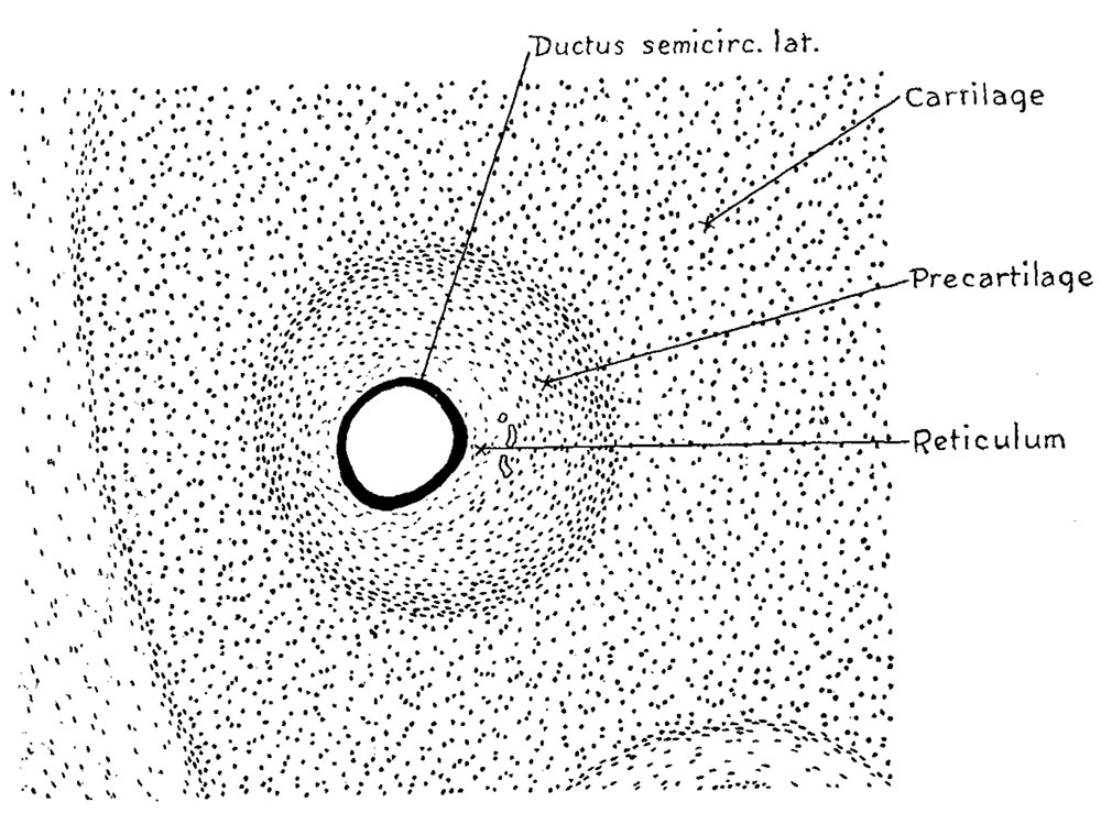
Fig. 7 Section passing transversely through the lateral semicircular canal in a human embryo 30 mm long
(Carnegie Collection No. 86), enlarged 100 diameters. The canal at this time is only slightly larger than the contained epithelial duct, but the zone of temporary precartilage marks out an area that is soon to be excavated by the process of dedifferentiation through which it becomes converted into a reticular connective tissue.
In the 30 mm embryo shown in figure 7, the first of these three figures, it will be seen that the epithelial duct is separated from the main cartilaginous mass of the capsule by a surrounding zone of precartilage and intervening between the latterand the duct is a narrow zone of mesenchymal tissue which is somewhat reticular in character. This zone of reticulum has attained its greatest width on the median side of the duct, toward the right, being at this point about twice as wide as the thickness of the duct wall. It is characterized by its reticular arrangement and by the presence of small blood vessels which are not found in the precartilage, although they lie closely against its inner margin. The area of precartilage stands out conspicuously in material that has been intensely stained in hematoxylin without any counterstain.
| Historic Disclaimer - information about historic embryology pages |
|---|
 |
- Links: Fig 1 | Fig 2 | Fig 3 | Fig 4 | Fig 5 | Fig 6 | Fig 7 | Fig 8 | Fig 9 | Fig 10 | Streeter 1917 | Historic Embryology Papers | Carnegie Embryos
Reference
Streeter GL. The factors involved in the excavation of the cavities in the cartilaginous capsule of the ear in the human embryo. (1917) Amer. J Anat. 22: 1–25.
Cite this page: Hill, M.A. (2024, April 27) Embryology Streeter1917 fig07.jpg. Retrieved from https://embryology.med.unsw.edu.au/embryology/index.php/File:Streeter1917_fig07.jpg
- © Dr Mark Hill 2024, UNSW Embryology ISBN: 978 0 7334 2609 4 - UNSW CRICOS Provider Code No. 00098G
File history
Click on a date/time to view the file as it appeared at that time.
| Date/Time | Thumbnail | Dimensions | User | Comment | |
|---|---|---|---|---|---|
| current | 09:36, 30 October 2015 | | 1,000 × 739 (198 KB) | Z8600021 (talk | contribs) | |
| 09:33, 30 October 2015 | 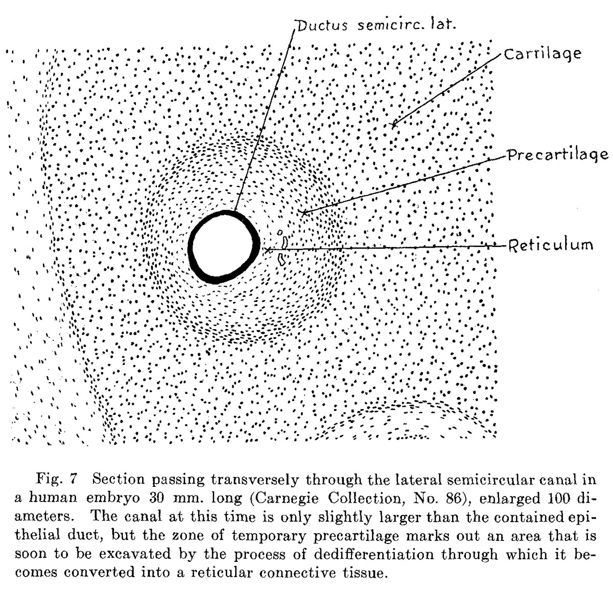 | 1,200 × 1,164 (337 KB) | Z8600021 (talk | contribs) | {{Streeter1917a}} Category:Carnegie Embryo 721 |
You cannot overwrite this file.
File usage
The following page uses this file: