File:Streeter1917 fig01.jpg: Difference between revisions
mNo edit summary |
mNo edit summary |
||
| (One intermediate revision by the same user not shown) | |||
| Line 1: | Line 1: | ||
==Fig. 1. Median views of wax-plate models of the left membranous labyrinth in human embryos== | ==Fig. 1. Median views of wax-plate models of the left membranous labyrinth in human embryos== | ||
Embryos have crown-rump lengths as indicated in the figure. The largest one is taken from Schonemann (’04) and represents the adult condition. They are all on the same scale of enlargement (4.4 diameters) and thus comparison of them shows graphically the amount of growth the labyrinth experiences during this period. | |||
The actual amount of increase in size of the labyrinth is graphically pictured in figure 1. The outlines are made so that they show on the same scale of enlargement a series of wax-plate models of the left membranous labyrinth of human embryos having a crown-rump length of 20, 30, 50, 85 and 130 mm., as indicated in the figure. This covers the periodiduring which the otic capsule is in a cartilaginous state. Ossification begins when the fetus has attained a crown-rump length of about 130 mm. | |||
{{Streeter1917a}} | {{Streeter1917a}} | ||
Latest revision as of 08:25, 30 October 2015
Fig. 1. Median views of wax-plate models of the left membranous labyrinth in human embryos
Embryos have crown-rump lengths as indicated in the figure. The largest one is taken from Schonemann (’04) and represents the adult condition. They are all on the same scale of enlargement (4.4 diameters) and thus comparison of them shows graphically the amount of growth the labyrinth experiences during this period.
The actual amount of increase in size of the labyrinth is graphically pictured in figure 1. The outlines are made so that they show on the same scale of enlargement a series of wax-plate models of the left membranous labyrinth of human embryos having a crown-rump length of 20, 30, 50, 85 and 130 mm., as indicated in the figure. This covers the periodiduring which the otic capsule is in a cartilaginous state. Ossification begins when the fetus has attained a crown-rump length of about 130 mm.
| Historic Disclaimer - information about historic embryology pages |
|---|
 |
- Links: Fig 1 | Fig 2 | Fig 3 | Fig 4 | Fig 5 | Fig 6 | Fig 7 | Fig 8 | Fig 9 | Fig 10 | Streeter 1917 | Historic Embryology Papers | Carnegie Embryos
Reference
Streeter GL. The factors involved in the excavation of the cavities in the cartilaginous capsule of the ear in the human embryo. (1917) Amer. J Anat. 22: 1–25.
Cite this page: Hill, M.A. (2024, May 3) Embryology Streeter1917 fig01.jpg. Retrieved from https://embryology.med.unsw.edu.au/embryology/index.php/File:Streeter1917_fig01.jpg
- © Dr Mark Hill 2024, UNSW Embryology ISBN: 978 0 7334 2609 4 - UNSW CRICOS Provider Code No. 00098G
File history
Click on a date/time to view the file as it appeared at that time.
| Date/Time | Thumbnail | Dimensions | User | Comment | |
|---|---|---|---|---|---|
| current | 08:10, 30 October 2015 | 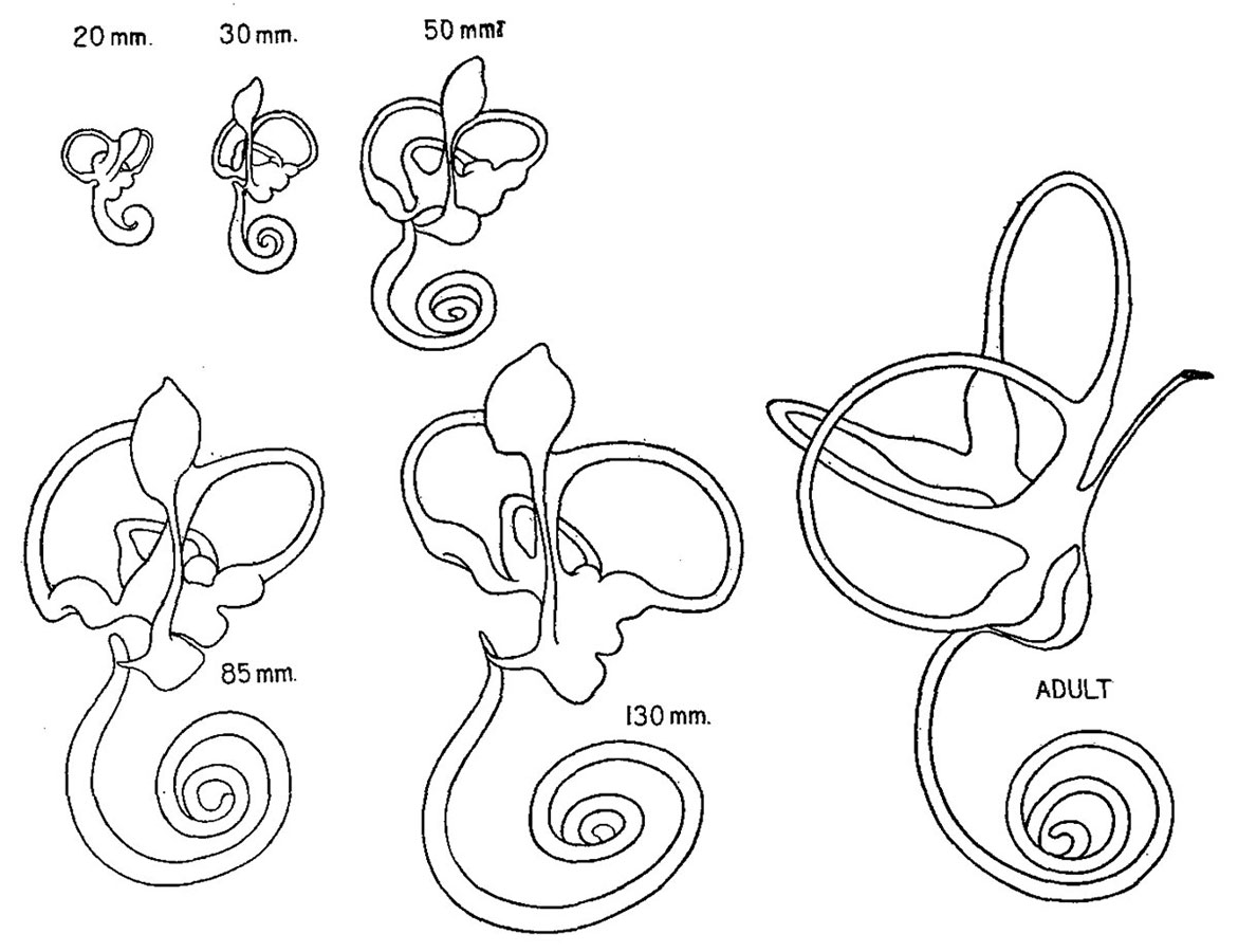 | 1,171 × 900 (131 KB) | Z8600021 (talk | contribs) | |
| 07:18, 30 October 2015 | 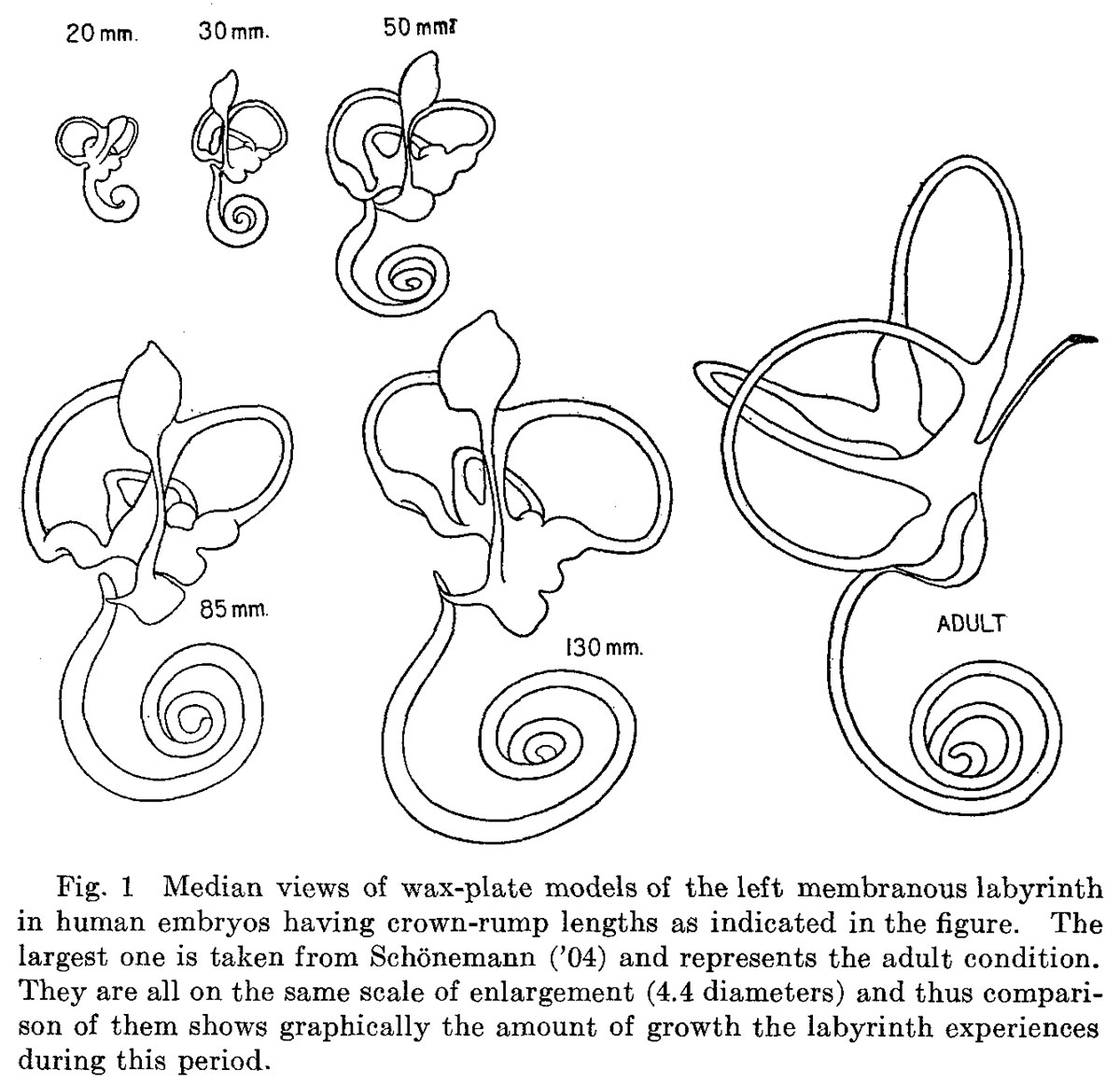 | 1,200 × 1,162 (223 KB) | Z8600021 (talk | contribs) |
You cannot overwrite this file.
File usage
The following page uses this file: