File:Streeter1917-fig09.jpg: Difference between revisions
mNo edit summary |
mNo edit summary |
||
| Line 1: | Line 1: | ||
==Figure 9. Medial Views of left membranous labyrinth human fetus 130 mm== | |||
Median View of the same model shown in figure 8. The oval impression on the proximal end of the scala tympani corresponds to the fenestra cochleae. As yet there is no communication at this point between the scala tympani and the subaraehnoid spaces corresponding to the aquaeductus cochleae. The spaces making up the cistern cover almost the whole of the utricle and saecule excepting the places at which the nerves enter and a small part of the medial surface near the attachment of the appendage. | |||
{{Streeter1917 figures}} | {{Streeter1917 figures}} | ||
Revision as of 14:40, 17 September 2015
Figure 9. Medial Views of left membranous labyrinth human fetus 130 mm
Median View of the same model shown in figure 8. The oval impression on the proximal end of the scala tympani corresponds to the fenestra cochleae. As yet there is no communication at this point between the scala tympani and the subaraehnoid spaces corresponding to the aquaeductus cochleae. The spaces making up the cistern cover almost the whole of the utricle and saecule excepting the places at which the nerves enter and a small part of the medial surface near the attachment of the appendage.
| Historic Disclaimer - information about historic embryology pages |
|---|
 |
Reference
Streeter GL. The development of the scala tympani, scala vestibuli and perioticular cistern in the human embryo. (1917) Amer. J Anat. 21: 300-320.
Cite this page: Hill, M.A. (2024, May 18) Embryology Streeter1917-fig09.jpg. Retrieved from https://embryology.med.unsw.edu.au/embryology/index.php/File:Streeter1917-fig09.jpg
- © Dr Mark Hill 2024, UNSW Embryology ISBN: 978 0 7334 2609 4 - UNSW CRICOS Provider Code No. 00098G
File history
Click on a date/time to view the file as it appeared at that time.
| Date/Time | Thumbnail | Dimensions | User | Comment | |
|---|---|---|---|---|---|
| current | 14:36, 17 September 2015 | 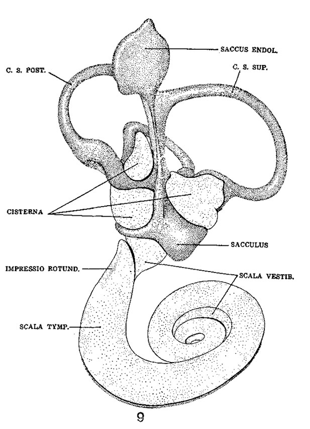 | 617 × 853 (109 KB) | Z8600021 (talk | contribs) | |
| 14:36, 17 September 2015 |  | 1,200 × 853 (218 KB) | Z8600021 (talk | contribs) | ||
| 14:35, 17 September 2015 | 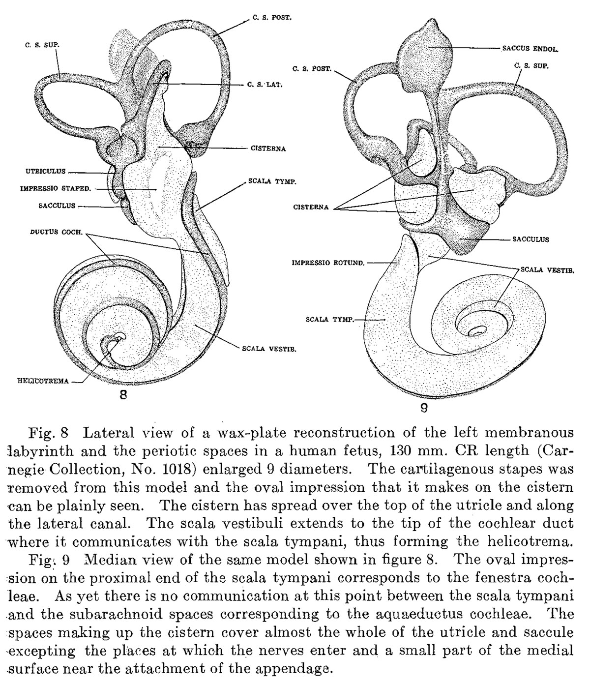 | 1,200 × 1,391 (409 KB) | Z8600021 (talk | contribs) |
You cannot overwrite this file.
File usage
The following page uses this file: