File:Streeter1917-fig02.jpg

Original file (1,000 × 847 pixels, file size: 276 KB, MIME type: image/jpeg)
==Fig. 2 Detail of the section shown in figure 1, enlarged 278 diameters. This figure shows the part of the cochlear duct that is to form the organ of Corti, and the adjacent tissue that becomes incorporated in the basilar membrane. Below this is the periotic reticulum whose spaces are in the process of enlarging. By repeated coalescence these spaces finally unite with the large space that constitutes the scala tympani. This figure shows the histological appearance of the reticulum where the formation of tissue spaces is in active operation.
File history
Click on a date/time to view the file as it appeared at that time.
| Date/Time | Thumbnail | Dimensions | User | Comment | |
|---|---|---|---|---|---|
| current | 13:19, 16 September 2015 | | 1,000 × 847 (276 KB) | Z8600021 (talk | contribs) | |
| 13:18, 16 September 2015 | 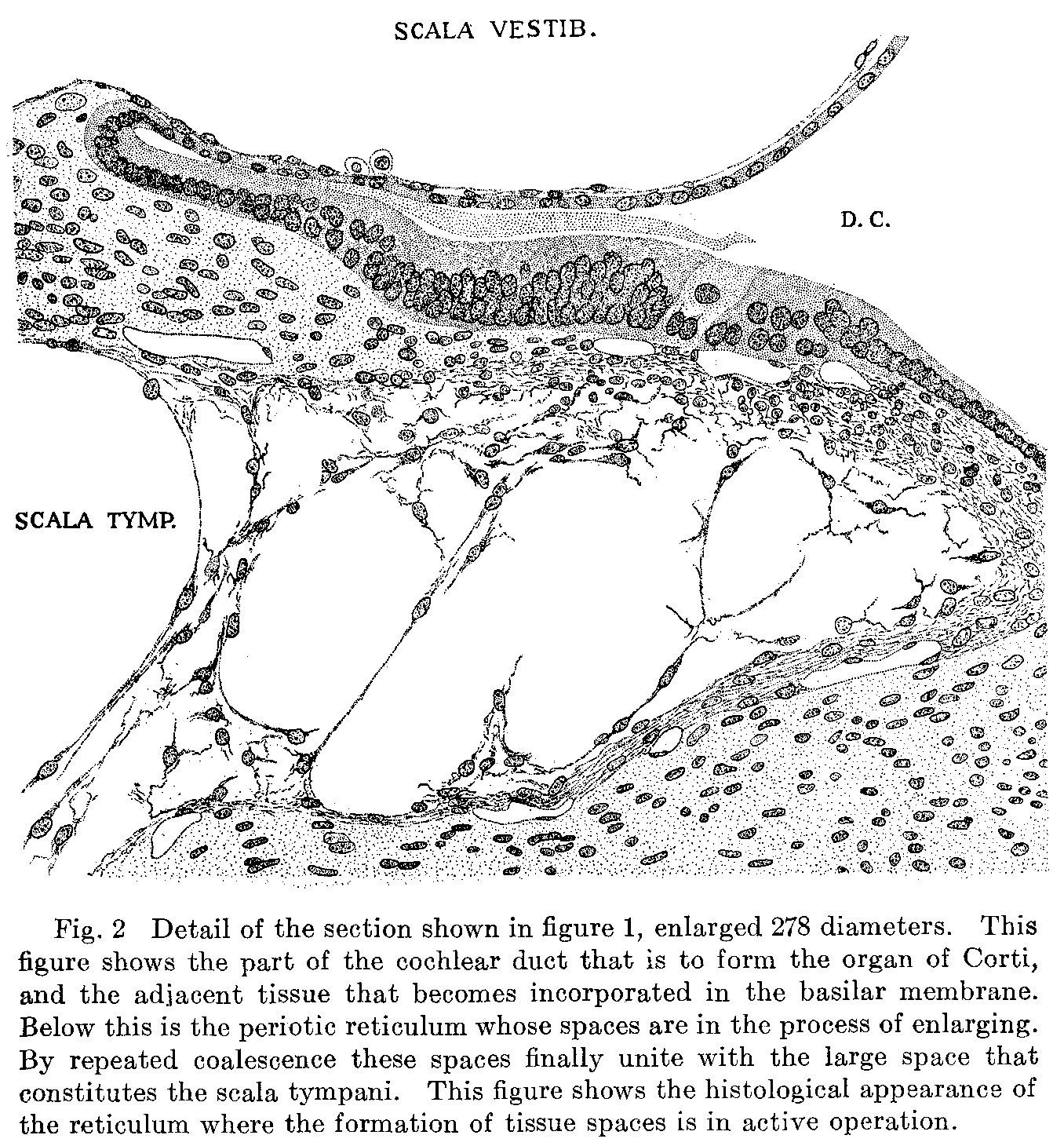 | 1,356 × 1,460 (646 KB) | Z8600021 (talk | contribs) | ==Fig. 2 Detail of the section shown in figure 1, enlarged 278 diameters. This figure shows the part of the cochlear duct that is to form the organ of Corti, and the adjacent tissue that becomes incorporated in the basilar membrane. Below this is the... |
You cannot overwrite this file.
File usage
There are no pages that use this file.