File:Streeter1915 fig04.jpg: Difference between revisions
(Z8600021 uploaded a new version of File:Streeter1915 fig04.jpg) |
mNo edit summary |
||
| Line 1: | Line 1: | ||
==Fig. 4 Profile reconstruction of the Veins of the dura mater in a human embryo 21 mm long== | |||
(No. 460, Carnegie Collection). The Vein marked ‘X’ is the remnant of the otic portion of the primary head vein which originally connected the cavernous region with the internal jugular vein. The outlines of the central nervous system in this model were taken from a model made by Professor Lewis. Enlarged 10 diameters. | |||
{{Historic Disclaimer}} | {{Historic Disclaimer}} | ||
| Line 8: | Line 13: | ||
{{Footer}} | {{Footer}} | ||
[[Category:Carnegie Embryo | [[Category:Carnegie Embryo 460]] | ||
Latest revision as of 10:46, 26 November 2016
Fig. 4 Profile reconstruction of the Veins of the dura mater in a human embryo 21 mm long
(No. 460, Carnegie Collection). The Vein marked ‘X’ is the remnant of the otic portion of the primary head vein which originally connected the cavernous region with the internal jugular vein. The outlines of the central nervous system in this model were taken from a model made by Professor Lewis. Enlarged 10 diameters.
| Historic Disclaimer - information about historic embryology pages |
|---|
 |
Links: fig 1 | fig 2 | fig 3 | fig 4 | fig 5 | fig 6 | fig 7 | fig 8 | fig 9 | fig 10 | fig 11 | fig 12 | fig 13 | fig 14 | fig 15 | fig 16 | fig 17 | Streeter 1915 | Neural Vascular | Historic Papers
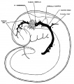
fig 1 embryo 4 mm No. 588
fig 2 embryo 13.8 mm No. 940
fig 3 embryo 18 mm No. 144

fig 4 embryo 21 mm No. 460
fig 5 embryo 24 mm No. 632
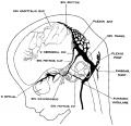
fig 6 embryo 50 mm No. 96
fig 7 embryo 13.8 mm No. 940
fig 8 embryo 20 mm No. 349
fig 9 fetus 54 mm No. 458
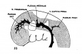
fig 10 embryo 4 mm No. 588
fig 11 embryo 14 mm No. 940
fig 12 embryo 18 mm No. 144
fig 13 embryo 21 mm No. 460
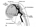
fig 14 embryo 35 mm No. 100
fig 15 embryo 50 mm No. 96
fig 16 embryo 80 mm No. 234a
fig 17 adult


Reference
Streeter GL. The development of the venous sinuses of the dura mater in the human embryo. (1915) Amer. J Anat.18: 145-178.
Cite this page: Hill, M.A. (2024, May 17) Embryology Streeter1915 fig04.jpg. Retrieved from https://embryology.med.unsw.edu.au/embryology/index.php/File:Streeter1915_fig04.jpg
- © Dr Mark Hill 2024, UNSW Embryology ISBN: 978 0 7334 2609 4 - UNSW CRICOS Provider Code No. 00098G
File history
Click on a date/time to view the file as it appeared at that time.
| Date/Time | Thumbnail | Dimensions | User | Comment | |
|---|---|---|---|---|---|
| current | 10:44, 26 November 2016 | 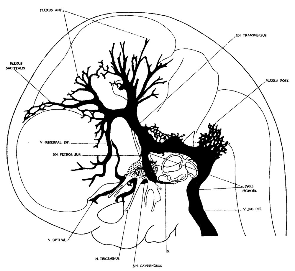 | 1,000 × 919 (130 KB) | Z8600021 (talk | contribs) | |
| 10:44, 26 November 2016 | 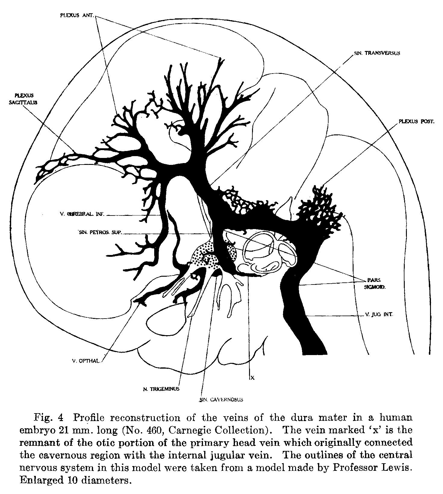 | 1,471 × 1,639 (346 KB) | Z8600021 (talk | contribs) | {{Historic Disclaimer}} {{Streeter1915 figures}} ===Reference=== {{Ref-Streeter1915}} {{Footer}} Category:Carnegie Embryo 458 |
You cannot overwrite this file.
File usage
The following 21 pages use this file:
- Neural - Meninges Development
- Paper - The development of the venous sinuses of the dura mater in the human embryo
- File:Streeter1915 fig01.jpg
- File:Streeter1915 fig02.jpg
- File:Streeter1915 fig03.jpg
- File:Streeter1915 fig04.jpg
- File:Streeter1915 fig05.jpg
- File:Streeter1915 fig06.jpg
- File:Streeter1915 fig07.jpg
- File:Streeter1915 fig08.jpg
- File:Streeter1915 fig09.jpg
- File:Streeter1915 fig10.jpg
- File:Streeter1915 fig11.jpg
- File:Streeter1915 fig12.jpg
- File:Streeter1915 fig13.jpg
- File:Streeter1915 fig14.jpg
- File:Streeter1915 fig15.jpg
- File:Streeter1915 fig16.jpg
- File:Streeter1915 fig17.jpg
- File:Streeter1915 table01.jpg
- Template:Streeter1915 figures