File:Streeter1908 fig02.jpg
From Embryology
Size of this preview: 800 × 313 pixels. Other resolution: 1,000 × 391 pixels.
Original file (1,000 × 391 pixels, file size: 39 KB, MIME type: image/jpeg)
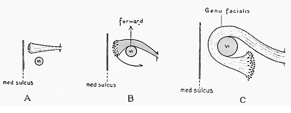
Fig. 2. Diagram illustrating the development of the genu of the facial nerve in the human embryo
The drawings show the right facial nerve and its nucleus of origin, in three stages : the youngest, A. being the 10 mm. embryo, and the oldest, C. the new-born child. The relative position of the nucleus of the Abducens nerve is represented in outline. Its nerve trunk could not be shown, as the structures are represented as seen from above.
File history
Click on a date/time to view the file as it appeared at that time.
| Date/Time | Thumbnail | Dimensions | User | Comment | |
|---|---|---|---|---|---|
| current | 23:31, 15 February 2016 | 1,000 × 391 (39 KB) | Z8600021 (talk | contribs) | ||
| 23:31, 15 February 2016 |  | 1,330 × 704 (146 KB) | Z8600021 (talk | contribs) |
You cannot overwrite this file.
File usage
The following 2 pages use this file: