File:Streeter1906 fig04.jpg: Difference between revisions
mNo edit summary |
mNo edit summary |
||
| Line 1: | Line 1: | ||
==Fig.4. Diagram representing the growth and stages of differentiation of the human membranous labyrinth== | ==Fig.4. Diagram representing the growth and stages of differentiation of the human membranous labyrinth== | ||
{| | {| | ||
| col width="150px"|3.5 weeks (6-7 mm.) the ear vesicle consists of two simple pouches, into the upper of which opens the endolymphatic appendage. | | col width="150px" valign=top|'''3.5 weeks''' (6-7 mm.) the ear vesicle consists of two simple pouches, into the upper of which opens the endolymphatic appendage. | ||
| col width=150px| | | col width=150px|'''4 weeks''' (9 mm.) there is at the base of the vestibular pouch an atrium, the space destined to form the utricle and saccule. | ||
| col width=150px| | | col width=150px|'''5 weeks''' (12 mm.) this space is circumscribed from the cochlear pouch below by a constriction corresponding to the ductus reuniens, and above from the rest of the vestibular pouch by the formation of the semicircular canals. | ||
| col width=150px| | | col width=150px|'''6 weeks''' (20 mm.) an ingrowth of the wall of the atrium divides it into an upper part (utricle) and lower part (saccule). | ||
| col width=150px| | | col width=150px|'''10 weeks''' (30 mm.) this partition between the utricle and saccule is complete and extends inward in such a way as to split the orifice of the endolymphatic duct. | ||
|} | |} | ||
Revision as of 17:59, 23 July 2015
Fig.4. Diagram representing the growth and stages of differentiation of the human membranous labyrinth
| 3.5 weeks (6-7 mm.) the ear vesicle consists of two simple pouches, into the upper of which opens the endolymphatic appendage. | 4 weeks (9 mm.) there is at the base of the vestibular pouch an atrium, the space destined to form the utricle and saccule. | 5 weeks (12 mm.) this space is circumscribed from the cochlear pouch below by a constriction corresponding to the ductus reuniens, and above from the rest of the vestibular pouch by the formation of the semicircular canals. | 6 weeks (20 mm.) an ingrowth of the wall of the atrium divides it into an upper part (utricle) and lower part (saccule). | 10 weeks (30 mm.) this partition between the utricle and saccule is complete and extends inward in such a way as to split the orifice of the endolymphatic duct. |
| Historic Disclaimer - information about historic embryology pages |
|---|
 |
- Mall 1906 Links: Fig 1. 14mm Embryo | Fig 2. 30mm Embryo | Fig 3. Semicircular canal | Fig 4. Membranous Labyrinth | Fig 5. Acoustic nerve complex | Fig 6. Facial-acoustic Complex | Fig 7. Facial Nerve Pig Embryo 20 cm | Fig 8. Geniculate Ganglion | Plate 1. Human Embryo 4 to 20 mm | Plate 2. Human Embryo 30 mm | Membranous Labyrinth and Nerves
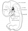
Fig 1 Membranous Labyrinth Human Embryo 14 mm
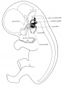
Fig 2 30mm Embryo
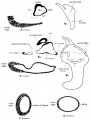
Fig 3 Semicircular canal
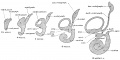
Fig 4 Membranous Labyrinth Growth
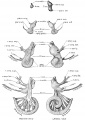
Fig 5 Acoustic nerve complex
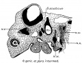
Fig 6 Facial-acoustic Complex Human Embryo 7 mm
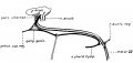
Fig 7 Facial Nerve Pig Embryo 20 cm
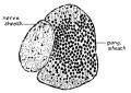
Fig 8 Geniculate Ganglion Human Embryo 30 mm
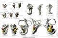
Plate 1. Membranous Labyrinth Human Embryo 4 to 20 mm
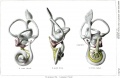
Plate 2. Membranous Labyrinth Human Embryo 30 mm


Reference
Streeter GL. On the development of the membranous labyrinth and the acoustic and facial nerves in the human embryo. (1906) Amer. J Anat. 6:139-165.
Cite this page: Hill, M.A. (2024, May 19) Embryology Streeter1906 fig04.jpg. Retrieved from https://embryology.med.unsw.edu.au/embryology/index.php/File:Streeter1906_fig04.jpg
- © Dr Mark Hill 2024, UNSW Embryology ISBN: 978 0 7334 2609 4 - UNSW CRICOS Provider Code No. 00098G
File history
Click on a date/time to view the file as it appeared at that time.
| Date/Time | Thumbnail | Dimensions | User | Comment | |
|---|---|---|---|---|---|
| current | 17:49, 23 July 2015 |  | 2,237 × 1,113 (404 KB) | Z8600021 (talk | contribs) | |
| 17:49, 23 July 2015 |  | 2,253 × 1,247 (618 KB) | Z8600021 (talk | contribs) |
You cannot overwrite this file.
File usage
The following 13 pages use this file:
- Hearing - Inner Ear Development
- Paper - On the development of the membranous labyrinth and the acoustic and facial nerves in the human embryo
- File:Streeter1906 fig01.jpg
- File:Streeter1906 fig02.jpg
- File:Streeter1906 fig03.jpg
- File:Streeter1906 fig04.jpg
- File:Streeter1906 fig05.jpg
- File:Streeter1906 fig06.jpg
- File:Streeter1906 fig07.jpg
- File:Streeter1906 fig08.jpg
- File:Streeter1906 plate01.jpg
- File:Streeter1906 plate02.jpg
- Template:Streeter1906 figures