File:Streeter028.jpg
Original file (774 × 1,000 pixels, file size: 69 KB, MIME type: image/jpeg)
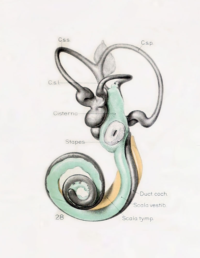
Fig. 28. Lateral view of left membranous labyrinth and the periotic spaces in a human fetus 85 mm CRL
Lateral view of wax-plate reconstruction of the left membranous labyrinth and the periotic spaces in a human fetus 85 mm. crown-rump length (Carnegie Collection, No. 1400-30), enlarged 11.4 diameters.
The cistern and the connecting scala vestibuli are shown in green. Although the greater part of the cistern abuts against the stapes, it will be noted that it is also begiiming to spread over the liorsal surface of the utricle and along the inner border of the lateral semicircular duct. The scala vestibule communicates freely with the cistern and extends downward alotig the apical surface of the cochlear duct, throughout nearly two turns, showing the characteristic sacculated appearance near its tip, where the coalescence of the spaces is less complete.
The figures shown on this plate 4 represent a series of median and lateral views of wax-plate reconstructions of the membranous labyrinth and the surrounding periotic tissue-spaces. They illustrate under the same scale of enlargement three typical stages in the development of these spaces.
Abbreviations
- C. s. 1. - ductus semicircuiaris lateralis
- C. s. p. - ductus semicircularis posterior
- C. s. s. - ductus semicircularis superior
- Duct, coch., = ductus cochlearis
- Impressio rotund. - area opposite the fenestra cochleae
- Impressio staped., area in contact with base of stapes
- Saccus endol. - saccus endolymphaticus
- Scala tymp. - scala tynipani
- Scala vestib. - scala vestibule.
Reference
Streeter G.L. The histogenesis and growth of the otic capsule and its contained periotic tissue-spaces in the human embryo Contributions to Embryology Carnegie Institution No.20 (1918) pp5-54, 4 text-figures and 4 plates.
File history
Click on a date/time to view the file as it appeared at that time.
| Date/Time | Thumbnail | Dimensions | User | Comment | |
|---|---|---|---|---|---|
| current | 21:51, 22 April 2012 | | 774 × 1,000 (69 KB) | Z8600021 (talk | contribs) | ====Fig. 28==== Lateral view of wax-plate reconstruction of the left membranous labyrinth and the periotic spaces in a human fetus 85 mm. crown-rump length (Carnegie Collection, No. 1400-30), enlarged 11.4 diameters. The cistern and the connecting scala v |
You cannot overwrite this file.
File usage
The following 3 pages use this file: