File:Streeter004.jpg
Original file (487 × 800 pixels, file size: 92 KB, MIME type: image/jpeg)
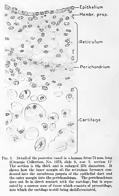
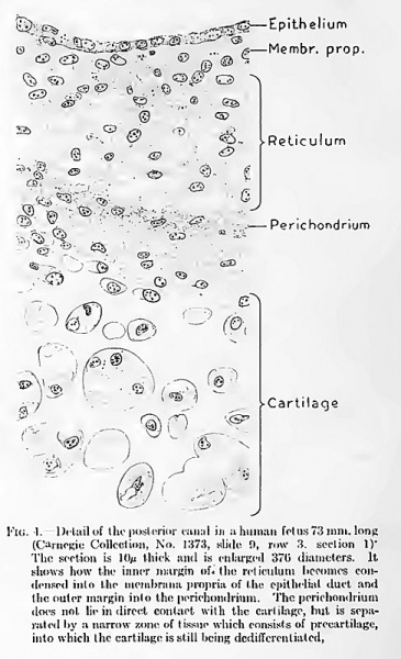
Fig. 4. Detail of the posterior canal in a human fetus 73 mm. long
(Carnegie Collection, No. 1373, slide 9, row 3. section 1) The section is 10 microns thick and is enlarged 370 diameters (in original printed version).
It shows how the inner margin of the reticulum becomes condensed into the membrane propria of the epithelial duct and the outer margin into the perichondrium.
The perichondrium does not lie in direct contact with the cartilage, but is separated by a narrow zone of tissue which consists of precartilage, into which the cartilage is still being dedifferentiated.
Reference
Streeter G.L. The histogenesis and growth of the otic capsule and its contained periotic tissue-spaces in the human embryo Contributions to Embryology Carnegie Institution No.20 (1918) pp5-54, 4 text-figures and 4 plates.
File history
Click on a date/time to view the file as it appeared at that time.
| Date/Time | Thumbnail | Dimensions | User | Comment | |
|---|---|---|---|---|---|
| current | 00:13, 15 February 2011 | | 487 × 800 (92 KB) | S8600021 (talk | contribs) |
You cannot overwrite this file.
File usage
The following 2 pages use this file: