File:Stewart1955 plate01.jpg: Difference between revisions
(Z8600021 uploaded a new version of File:Stewart1955 plate01.jpg) |
mNo edit summary |
||
| Line 1: | Line 1: | ||
==Plate 1== | |||
Represents the left half of the brain of a human embryo of 15.0 mm, C-R length with an estimated ovulation age of 37 days. It is a graphic reconstruction of the configurations of the nuclei forming the medial wall of the mesencephalon and diencephalon and the medial external wall of the anterior portion of the left cerebral hemisphere. This graphic reconstruction was done to provide a basis to which the series of embryos could be related, and was constructed ty serial cross sections. The term nucleus as used in these descriptions does not necessarily indicate a region directly comparable to a nucleus in the adult sense, but indicates an area of cellular concentration which can be identified in the forebrain of successively older embryos. The names given to the tracts and nuclei have been based on the names of corresponding nuclei and tracts in the adult brain wherever it has been possible to identilNr the embryonic structure with its adult derivitive. | |||
===Reference=== | ===Reference=== | ||
Revision as of 09:38, 11 June 2018
Plate 1
Represents the left half of the brain of a human embryo of 15.0 mm, C-R length with an estimated ovulation age of 37 days. It is a graphic reconstruction of the configurations of the nuclei forming the medial wall of the mesencephalon and diencephalon and the medial external wall of the anterior portion of the left cerebral hemisphere. This graphic reconstruction was done to provide a basis to which the series of embryos could be related, and was constructed ty serial cross sections. The term nucleus as used in these descriptions does not necessarily indicate a region directly comparable to a nucleus in the adult sense, but indicates an area of cellular concentration which can be identified in the forebrain of successively older embryos. The names given to the tracts and nuclei have been based on the names of corresponding nuclei and tracts in the adult brain wherever it has been possible to identilNr the embryonic structure with its adult derivitive.
Reference
Stewart GG. The development of the blood supply to the human embryo basal ganglia. (1955) University of Alberta, Canada.
Cite this page: Hill, M.A. (2024, May 1) Embryology Stewart1955 plate01.jpg. Retrieved from https://embryology.med.unsw.edu.au/embryology/index.php/File:Stewart1955_plate01.jpg
- © Dr Mark Hill 2024, UNSW Embryology ISBN: 978 0 7334 2609 4 - UNSW CRICOS Provider Code No. 00098G
File history
Click on a date/time to view the file as it appeared at that time.
| Date/Time | Thumbnail | Dimensions | User | Comment | |
|---|---|---|---|---|---|
| current | 18:38, 10 June 2018 | 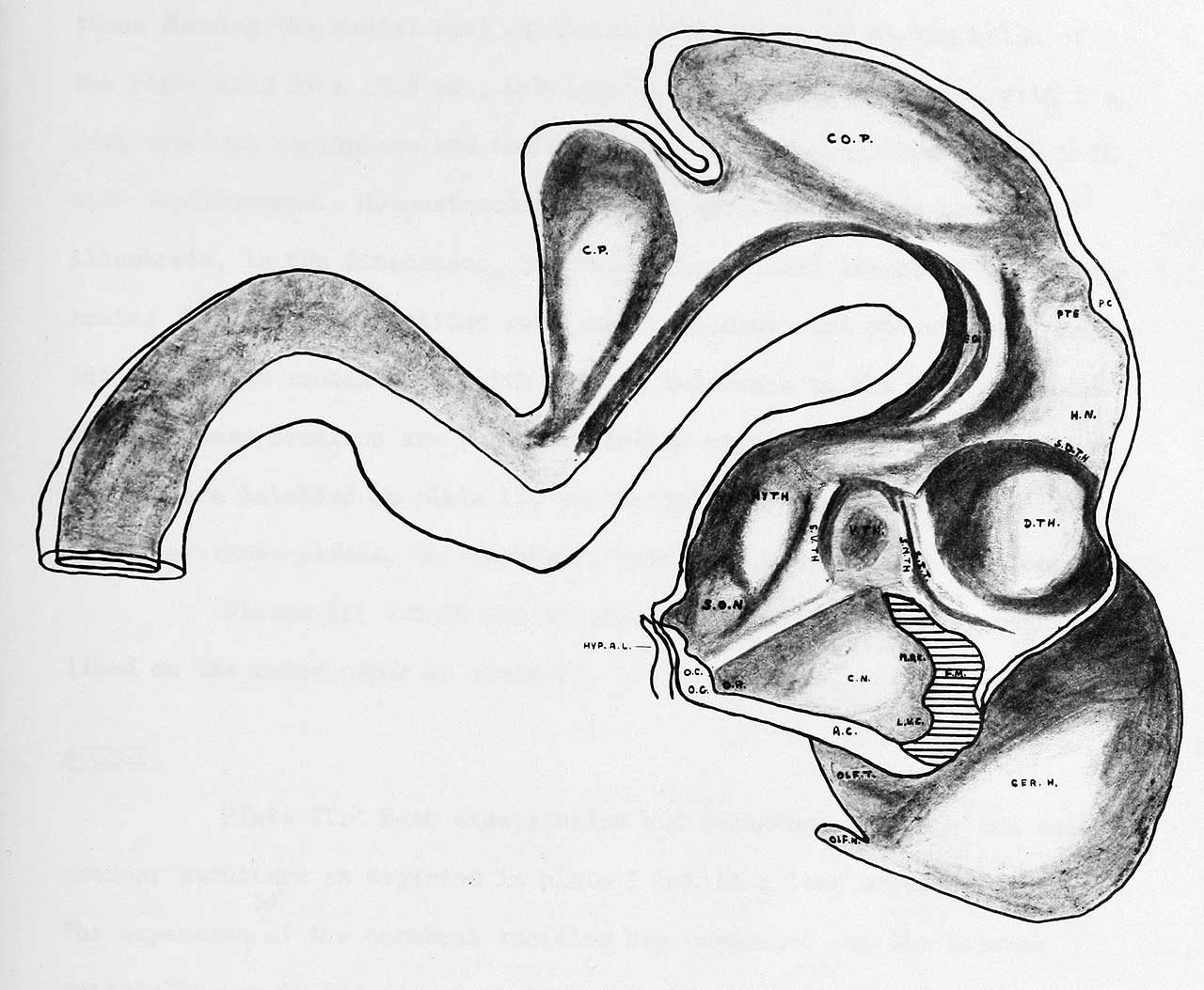 | 1,280 × 1,053 (135 KB) | Z8600021 (talk | contribs) | |
| 18:38, 10 June 2018 |  | 3,047 × 3,196 (673 KB) | Z8600021 (talk | contribs) |
You cannot overwrite this file.
File usage
The following page uses this file: