File:Stewart1955 frontispiece.jpg
From Embryology

Size of this preview: 521 × 599 pixels. Other resolution: 1,028 × 1,182 pixels.
Original file (1,028 × 1,182 pixels, file size: 131 KB, MIME type: image/jpeg)
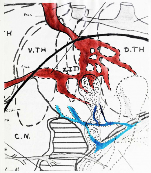
Frontispiece
(H51, original 40X)»
Showing the formation of the choroid plexus from the choroidal arteries at the 15.5 mm stage
Reference
Stewart GG. The development of the blood supply to the human embryo basal ganglia. (1955) University of Alberta, Canada.
Cite this page: Hill, M.A. (2024, May 17) Embryology Stewart1955 frontispiece.jpg. Retrieved from https://embryology.med.unsw.edu.au/embryology/index.php/File:Stewart1955_frontispiece.jpg
- © Dr Mark Hill 2024, UNSW Embryology ISBN: 978 0 7334 2609 4 - UNSW CRICOS Provider Code No. 00098G
File history
Click on a date/time to view the file as it appeared at that time.
| Date/Time | Thumbnail | Dimensions | User | Comment | |
|---|---|---|---|---|---|
| current | 18:00, 10 June 2018 | | 1,028 × 1,182 (131 KB) | Z8600021 (talk | contribs) | |
| 17:59, 10 June 2018 | Error creating thumbnail: File with dimensions greater than 12.5 MP | 3,237 × 4,419 (1.04 MB) | Z8600021 (talk | contribs) | ==Frontispiece== (H51, original 40X)» Showing the formation of the choroid plexus from the choroidal arteries at the 15.5 mm stage ===Reference=== {{Ref-Stewart1955}} |
You cannot overwrite this file.
File usage
The following page uses this file: