File:Stage 12 drosophila.jpg: Difference between revisions
From Embryology
No edit summary |
No edit summary |
||
| Line 2: | Line 2: | ||
Image courtesy of Katrin Weigmann, Robert Klapper, Thomas Strasser, Christof Rickert, Gerd Technau, Herbert Jäckle, Wilfried Janning and Christian Klämbt: FlyMove – a new way to look at development of Drosophila.Trends Genet. In press. http://flymove.uni-muenster.de Image used with permission from Christian Klämbt | Image courtesy of Katrin Weigmann, Robert Klapper, Thomas Strasser, Christof Rickert, Gerd Technau, Herbert Jäckle, Wilfried Janning and Christian Klämbt: FlyMove – a new way to look at development of Drosophila.Trends Genet. In press. http://flymove.uni-muenster.de Image used with permission from Christian Klämbt | ||
[[Category:Fly]] | |||
Latest revision as of 21:51, 10 September 2012
Stage 12 Drosophila embryo. The section is stained using an anti-Crumbs antibody, showing epithelial structures.
Image courtesy of Katrin Weigmann, Robert Klapper, Thomas Strasser, Christof Rickert, Gerd Technau, Herbert Jäckle, Wilfried Janning and Christian Klämbt: FlyMove – a new way to look at development of Drosophila.Trends Genet. In press. http://flymove.uni-muenster.de Image used with permission from Christian Klämbt
File history
Click on a date/time to view the file as it appeared at that time.
| Date/Time | Thumbnail | Dimensions | User | Comment | |
|---|---|---|---|---|---|
| current | 12:38, 18 September 2009 | 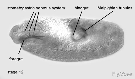 | 461 × 270 (22 KB) | Z3217015 (talk | contribs) | Stage 12 Drosophila embryo. The section is stained using an anti-Crumbs antibody, showing epithelial structures. Image courtesy of Katrin Weigmann, Robert Klapper, Thomas Strasser, Christof Rickert, Gerd Technau, Herbert Jäckle, Wilfried Janning and Chr |
You cannot overwrite this file.
File usage
The following page uses this file: