File:Stage8 nodal cilia.jpg: Difference between revisions
From Embryology
No edit summary |
No edit summary |
||
| Line 4: | Line 4: | ||
scanning electron micrograph (original file name [http://embryology.med.unsw.edu.au/wwwhuman/Stages/Images/SEMimages450/presomiteSt8d18ventralnode1.jpg presomiteSt8d18ventralnode1.jpg]) | scanning electron micrograph (original file name [http://embryology.med.unsw.edu.au/wwwhuman/Stages/Images/SEMimages450/presomiteSt8d18ventralnode1.jpg presomiteSt8d18ventralnode1.jpg]) | ||
'''Image Source:''' Prof Kathy Sulik scanning electron micrographs of the Carnegie stages of the early human embryo. No reproduction without permission. | '''Image Source:''' Prof Kathy Sulik scanning electron micrographs of the Carnegie stages of the early human embryo. No reproduction without permission. | ||
Revision as of 11:34, 14 August 2009
Human embryo (Carnegie stage 8)
This image is a high power selected region at the primitive node showing the cilia that are thought to rotate in one direction leading to a gradient of factors establishing the early left/right axis of the embryo.
scanning electron micrograph (original file name presomiteSt8d18ventralnode1.jpg)
Image Source: Prof Kathy Sulik scanning electron micrographs of the Carnegie stages of the early human embryo. No reproduction without permission.
File history
Click on a date/time to view the file as it appeared at that time.
| Date/Time | Thumbnail | Dimensions | User | Comment | |
|---|---|---|---|---|---|
| current | 15:36, 3 August 2009 | 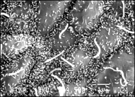 | 450 × 321 (50 KB) | MarkHill (talk | contribs) | Human embryo (Carnegie stage 8) This image is a high power selected region at the primitive node showing the cilia that are thought to rotate in one direction leading to a gradient of factors establishing the early left/right axis of the embryo. scannin |
You cannot overwrite this file.
File usage
The following 3 pages use this file: