File:Stage7-sem1.jpg: Difference between revisions
(Human Embryo Carnegie stage 7, 17 days, pre-somite, scanning electron micrograph image Embryonic disc (epiblast/ectoderm layer) dorsal view, with amniotic membrane partially removed. Primitive node (Henson's node) in centre of disc and primitive streak) |
No edit summary |
||
| Line 6: | Line 6: | ||
Primitive node (Henson's node) in centre of disc and primitive streak, shown as indentation in disc is extending to the left. Connecting stalk to the left. | Primitive node (Henson's node) in centre of disc and primitive streak, shown as indentation in disc is extending to the left. Connecting stalk to the left. | ||
See also [[:File:Stage7-sem2.jpg|Stage7-sem2.jpg]] where this image has been rotated 90 degrees counterclockwise. | |||
{{Template:SEM}} | {{Template:SEM}} | ||
[[Category:Human Embryo]] [[Category:Carnegie Stage]] [[Category:Week 3]] [[Category:Gastrulation]] | [[Category:Human Embryo]] [[Category:Carnegie Stage]] [[Category:Week 3]] [[Category:Gastrulation]] | ||
Revision as of 13:13, 21 August 2009
Human Embryo
Carnegie stage 7, 17 days, pre-somite, scanning electron micrograph image
Embryonic disc (epiblast/ectoderm layer) dorsal view, with amniotic membrane partially removed.
Primitive node (Henson's node) in centre of disc and primitive streak, shown as indentation in disc is extending to the left. Connecting stalk to the left.
See also Stage7-sem2.jpg where this image has been rotated 90 degrees counterclockwise.
Image Source: Scanning electron micrographs of the Carnegie stages of the early human embryos are reproduced with the permission of Prof Kathy Sulik, from embryos collected by Dr. Vekemans and Tania Attié-Bitach. Images are for educational purposes only and cannot be reproduced electronically or in writing without permission.
File history
Click on a date/time to view the file as it appeared at that time.
| Date/Time | Thumbnail | Dimensions | User | Comment | |
|---|---|---|---|---|---|
| current | 13:09, 21 August 2009 | 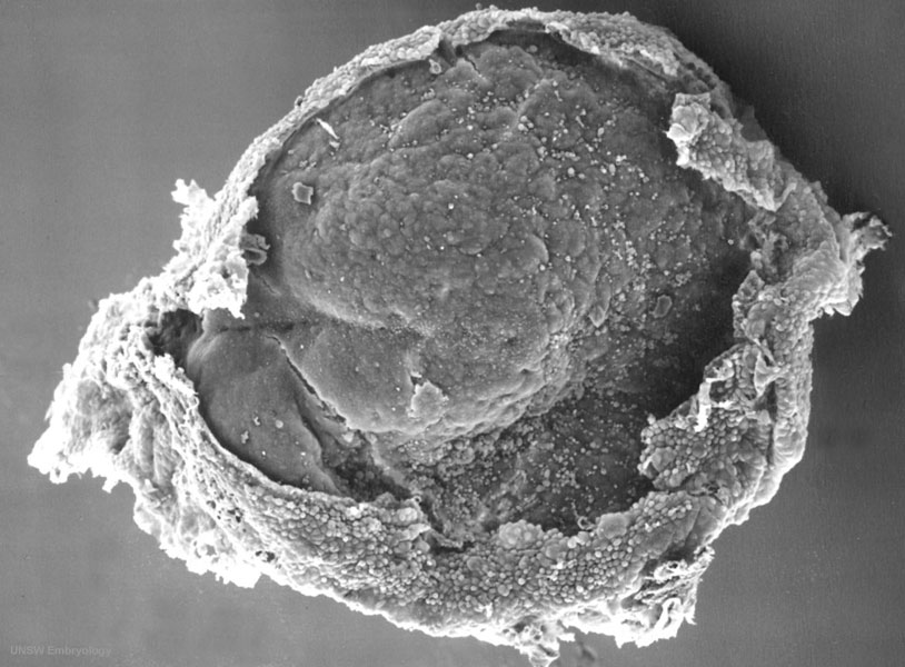 | 814 × 600 (100 KB) | MarkHill (talk | contribs) | Human Embryo Carnegie stage 7, 17 days, pre-somite, scanning electron micrograph image Embryonic disc (epiblast/ectoderm layer) dorsal view, with amniotic membrane partially removed. Primitive node (Henson's node) in centre of disc and primitive streak |
You cannot overwrite this file.
File usage
The following 5 pages use this file: