File:Stage12 sem9a cloacal membrane.jpg
Original file (719 × 1,000 pixels, file size: 115 KB, MIME type: image/jpeg)
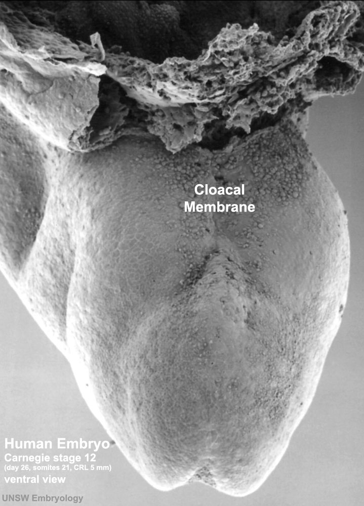
Human Embryo Carnegie Stage 12
A ventral caudal view of the embryo showing the region of the cloacal membrane and caudal neuropore.
Facts: Week 4, 26 days, 5 mm, Somite Number 25
View: Dorsolatera view, day 26, 25 somites, amniotic membrane removed
Features: caudal (dorsal) neuropore region just visible
Bright field image version of this image also available.
Original File Name: Stage12day26somite25 dorsal neuropore sem3.jpg
Image version links
Large 1200px | 1000px | Unlabeled 1200px | Unlabeled 1000px |
Related Images: Unlabeled version | 1000px |
- Stage 12 SEM Images: Bright Field 1 | Bright Field 3 | Bright Field 3 | SEM1 | SEM2 | SEM3 | SEM4 dorsolateral head and arches | SEM5 lateral head and arches | SEM6 ventrolateral head and arches | SEM7 lateral | SEM8 ventrolateral | SEM9 cloacal membrane | SEM9 labeled | Carnegie stage 12
Image Source: Scanning electron micrographs of the Carnegie stages of the early human embryos are reproduced with the permission of Prof Kathy Sulik, from embryos collected by Dr. Vekemans and Tania Attié-Bitach. Images are for educational purposes only and cannot be reproduced electronically or in writing without permission.
File history
Click on a date/time to view the file as it appeared at that time.
| Date/Time | Thumbnail | Dimensions | User | Comment | |
|---|---|---|---|---|---|
| current | 22:22, 29 May 2011 | | 719 × 1,000 (115 KB) | S8600021 (talk | contribs) | |
| 22:06, 29 May 2011 | 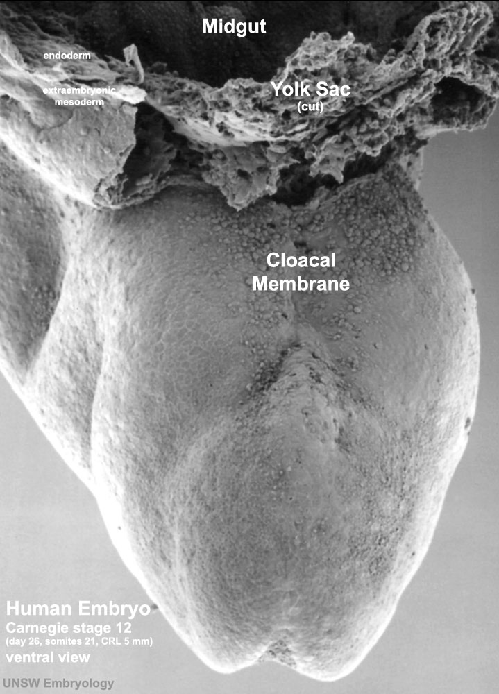 | 719 × 1,000 (118 KB) | S8600021 (talk | contribs) | == Human Embryo Carnegie Stage 12== A ventral caudal view of the embryo showing the region of the cloacal membrane and caudal neuropore. Facts: Week 4, 26 days, 5 mm, Somite Number 25 View: Dorsolatera view, day 26, 25 somites, amniotic membrane remove |
You cannot overwrite this file.
File usage
The following page uses this file: