File:Spaulding01.jpg
From Embryology
Size of this preview: 800 × 475 pixels. Other resolution: 1,045 × 621 pixels.
Original file (1,045 × 621 pixels, file size: 111 KB, MIME type: image/jpeg)
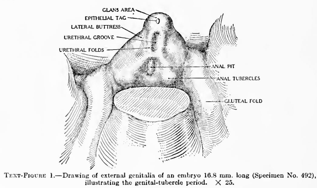
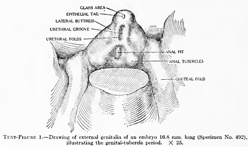
Figure 1. Drawing of external genitalia of an embryo 16.8 mm. long
(Specimen No. 492), illustrating the genital-tubercle period. X 25.
| Historic Disclaimer - information about historic embryology pages |
|---|
 |
Reference
The development of the external genitalia in the human embryo By Milo Herrick Spaulding, Of the University of Montmxa, Stale College of Agriculture, Bozeman. With four plates and two text-figures.
File history
Click on a date/time to view the file as it appeared at that time.
| Date/Time | Thumbnail | Dimensions | User | Comment | |
|---|---|---|---|---|---|
| current | 16:36, 26 March 2011 | | 1,045 × 621 (111 KB) | S8600021 (talk | contribs) | ==Figure 1. Drawing of external genitalia of an embryo 16.8 mm. long== (Specimen No. 492), illustrating the genital-tubercle period. X 25. {{Template:Historic Disclaimer}} ==Reference== The development of the external genitalia in the human embryo By |
You cannot overwrite this file.
File usage
The following 3 pages use this file: