File:Skull CT abnormal 06.jpg: Difference between revisions
No edit summary |
No edit summary |
||
| Line 3: | Line 3: | ||
Posterior (A), superior (B) and endocranial (C) 3DCT volume rendered images. There is no identifiable synostosis. The skull shape resembles a parallelogram (B), and the posterior skull base is not abnormally tilted (A). In addition, the posterior skull base axis coincides with the anterior skull base axis (C) | Posterior (A), superior (B) and endocranial (C) 3DCT volume rendered images. There is no identifiable synostosis. The skull shape resembles a parallelogram (B), and the posterior skull base is not abnormally tilted (A). In addition, the posterior skull base axis coincides with the anterior skull base axis (C) | ||
===Reference=== | |||
<pubmed>21431034</pubmed>| [http://www.ncbi.nlm.nih.gov/pmc/articles/PMC3056371 PMC3056371] | [http://www.ijri.org/article.asp?issn=0971-3026;year=2011;volume=21;issue=1;spage=49;epage=56;aulast=Khanna Indian J Radiol Imaging.] | |||
This is an open-access article distributed under the terms of the Creative Commons Attribution License, which permits unrestricted use, distribution, and reproduction in any medium, provided the original work is properly cited. | |||
Paritosh C Khanna © 2007 - 2012 Indian Journal of Radiology and Imaging | |||
Attribution-NonCommercial-ShareAlike 3.0 Unported ([http://creativecommons.org/licenses/by-nc-sa/3.0/ CC BY-NC-SA 3.0]) | |||
Original file name: Figure 7 Original figure has been modified, resized and relabelled. | |||
http://www.ijri.org/viewimage.asp?img=IndianJRadiolImaging_2011_21_1_49_76055_f8.jpg | http://www.ijri.org/viewimage.asp?img=IndianJRadiolImaging_2011_21_1_49_76055_f8.jpg | ||
[[Category:Human]] [[Category:Skull]] [[Category:Abnormal Development]] [[Category:Computed Tomography]] | |||
Revision as of 17:02, 17 March 2012
Deformational plagiocepahly
Posterior (A), superior (B) and endocranial (C) 3DCT volume rendered images. There is no identifiable synostosis. The skull shape resembles a parallelogram (B), and the posterior skull base is not abnormally tilted (A). In addition, the posterior skull base axis coincides with the anterior skull base axis (C)
Reference
<pubmed>21431034</pubmed>| PMC3056371 | Indian J Radiol Imaging.
This is an open-access article distributed under the terms of the Creative Commons Attribution License, which permits unrestricted use, distribution, and reproduction in any medium, provided the original work is properly cited.
Paritosh C Khanna © 2007 - 2012 Indian Journal of Radiology and Imaging
Attribution-NonCommercial-ShareAlike 3.0 Unported (CC BY-NC-SA 3.0)
Original file name: Figure 7 Original figure has been modified, resized and relabelled.
http://www.ijri.org/viewimage.asp?img=IndianJRadiolImaging_2011_21_1_49_76055_f8.jpg
File history
Click on a date/time to view the file as it appeared at that time.
| Date/Time | Thumbnail | Dimensions | User | Comment | |
|---|---|---|---|---|---|
| current | 16:59, 17 March 2012 | 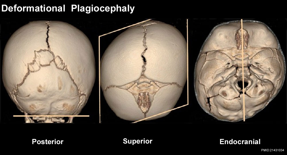 | 1,000 × 541 (85 KB) | Z8600021 (talk | contribs) |
You cannot overwrite this file.
File usage
The following 3 pages use this file: