File:Skull CT abnormal 05.jpg: Difference between revisions
No edit summary |
No edit summary |
||
| Line 2: | Line 2: | ||
Posterior (A), superior (B) and endocranial (C) 3DCT volume rendered images show posterior plagiocephaly secondary to unilateral lambdoid fusion (arrow). The posterior skull base is tilted downward on the affected side (A), and the skull assumes a trapezoid shape (B). Note also that the posterior skull base axis (passing through the basion and opisthion) is rotated toward the side of lambdoid fusion and does not coincide with the anterior skull base axis (passing through the crista galli and basion) (C) | Posterior (A), superior (B) and endocranial (C) 3DCT volume rendered images show posterior plagiocephaly secondary to unilateral lambdoid fusion (arrow). The posterior skull base is tilted downward on the affected side (A), and the skull assumes a trapezoid shape (B). Note also that the posterior skull base axis (passing through the basion and opisthion) is rotated toward the side of lambdoid fusion and does not coincide with the anterior skull base axis (passing through the crista galli and basion) (C) | ||
===Reference=== | |||
<pubmed>21431034</pubmed>| [http://www.ncbi.nlm.nih.gov/pmc/articles/PMC3056371 PMC3056371] | [http://www.ijri.org/article.asp?issn=0971-3026;year=2011;volume=21;issue=1;spage=49;epage=56;aulast=Khanna Indian J Radiol Imaging.] | |||
This is an open-access article distributed under the terms of the Creative Commons Attribution License, which permits unrestricted use, distribution, and reproduction in any medium, provided the original work is properly cited. | |||
Paritosh C Khanna © 2007 - 2012 Indian Journal of Radiology and Imaging | |||
Attribution-NonCommercial-ShareAlike 3.0 Unported ([http://creativecommons.org/licenses/by-nc-sa/3.0/ CC BY-NC-SA 3.0]) | |||
Original file name: Figure 6 Original figure has been modified, resized and relabelled. | |||
http://www.ijri.org/viewimage.asp?img=IndianJRadiolImaging_2011_21_1_49_76055_f7.jpg | http://www.ijri.org/viewimage.asp?img=IndianJRadiolImaging_2011_21_1_49_76055_f7.jpg | ||
[[Category:Human]] [[Category:Skull]] [[Category:Abnormal Development]] [[Category:Computed Tomography]] | |||
Revision as of 17:01, 17 March 2012
Posterior plagiocephaly
Posterior (A), superior (B) and endocranial (C) 3DCT volume rendered images show posterior plagiocephaly secondary to unilateral lambdoid fusion (arrow). The posterior skull base is tilted downward on the affected side (A), and the skull assumes a trapezoid shape (B). Note also that the posterior skull base axis (passing through the basion and opisthion) is rotated toward the side of lambdoid fusion and does not coincide with the anterior skull base axis (passing through the crista galli and basion) (C)
Reference
<pubmed>21431034</pubmed>| PMC3056371 | Indian J Radiol Imaging.
This is an open-access article distributed under the terms of the Creative Commons Attribution License, which permits unrestricted use, distribution, and reproduction in any medium, provided the original work is properly cited.
Paritosh C Khanna © 2007 - 2012 Indian Journal of Radiology and Imaging
Attribution-NonCommercial-ShareAlike 3.0 Unported (CC BY-NC-SA 3.0)
Original file name: Figure 6 Original figure has been modified, resized and relabelled.
http://www.ijri.org/viewimage.asp?img=IndianJRadiolImaging_2011_21_1_49_76055_f7.jpg
File history
Click on a date/time to view the file as it appeared at that time.
| Date/Time | Thumbnail | Dimensions | User | Comment | |
|---|---|---|---|---|---|
| current | 16:59, 17 March 2012 | 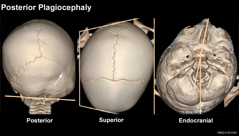 | 1,000 × 572 (88 KB) | Z8600021 (talk | contribs) |
You cannot overwrite this file.
File usage
The following 3 pages use this file: