File:Sinus venosus atrial septal defect 01.jpg: Difference between revisions
(http://www.annalspc.com/article.asp?issn=0974-2069;year=2014;volume=7;issue=2;spage=160;epage=162;aulast=Ganigara The role of cardiac MRI in the diagnosis and management of sinus venosus atrial septal defect. Ann Pediatr Cardiol. 2014 May;7(2):160-...) |
mNo edit summary |
||
| Line 15: | Line 15: | ||
Atrial septal defect; adult congenital heart disease; cardiac MRI | Atrial septal defect; adult congenital heart disease; cardiac MRI | ||
PMID 24987269 | PMID 24987269 | ||
Figure 1: (a) Breath-held fat suppressed three-dimensional steady-state free precession (SSFP) pulse sequence in diastole in the sagittal view demonstrating sinus venosus atrial septal defect (SV-ASD) (arrow) between superior vena cava (SVC) and left atrium (LA). (b) Breath-held fat suppressed three-dimensional SSFP pulse sequence in diastole in the axial view demonstrating SV-ASD (arrow) between SVC and LA. (c) Turbo spin-echo black blood image in the same axial plane as ure 1b demonstrating SV-ASD (arrow) between SVC and LA. (d) SSFP image showing the dilated right ventricle (RV) and left ventricle (LV) | |||
Revision as of 09:27, 22 February 2015
The role of cardiac MRI in the diagnosis and management of sinus venosus atrial septal defect.
Ann Pediatr Cardiol. 2014 May;7(2):160-2. doi: 10.4103/0974-2069.132509.
Ganigara M, Tanous D, Celermajer D, Puranik R.
Abstract
Sinus venosus atrial septal defects (SV-ASDs) are inter-atrial communications caused by a deficiency of the common wall between the superior or inferior vena cava and the right-sided pulmonary veins. The diagnosis can be challenging, especially in adults with delayed presentation. We present images that illustrate an example of the role of cardiac magnetic resonance imaging (CMRI) in the diagnosis and follow-up of a patient with SV-ASD. KEYWORDS: Atrial septal defect; adult congenital heart disease; cardiac MRI PMID 24987269
Figure 1: (a) Breath-held fat suppressed three-dimensional steady-state free precession (SSFP) pulse sequence in diastole in the sagittal view demonstrating sinus venosus atrial septal defect (SV-ASD) (arrow) between superior vena cava (SVC) and left atrium (LA). (b) Breath-held fat suppressed three-dimensional SSFP pulse sequence in diastole in the axial view demonstrating SV-ASD (arrow) between SVC and LA. (c) Turbo spin-echo black blood image in the same axial plane as ure 1b demonstrating SV-ASD (arrow) between SVC and LA. (d) SSFP image showing the dilated right ventricle (RV) and left ventricle (LV)
File history
Click on a date/time to view the file as it appeared at that time.
| Date/Time | Thumbnail | Dimensions | User | Comment | |
|---|---|---|---|---|---|
| current | 09:27, 22 February 2015 | 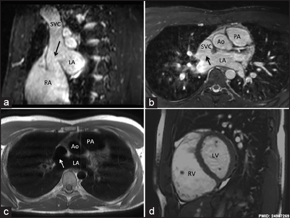 | 1,000 × 754 (90 KB) | Z8600021 (talk | contribs) | http://www.annalspc.com/article.asp?issn=0974-2069;year=2014;volume=7;issue=2;spage=160;epage=162;aulast=Ganigara The role of cardiac MRI in the diagnosis and management of sinus venosus atrial septal defect. Ann Pediatr Cardiol. 2014 May;7(2):160-... |
You cannot overwrite this file.
File usage
There are no pages that use this file.