File:Shaner1945 plate03.jpg
Original file (1,280 × 1,977 pixels, file size: 475 KB, MIME type: image/jpeg)
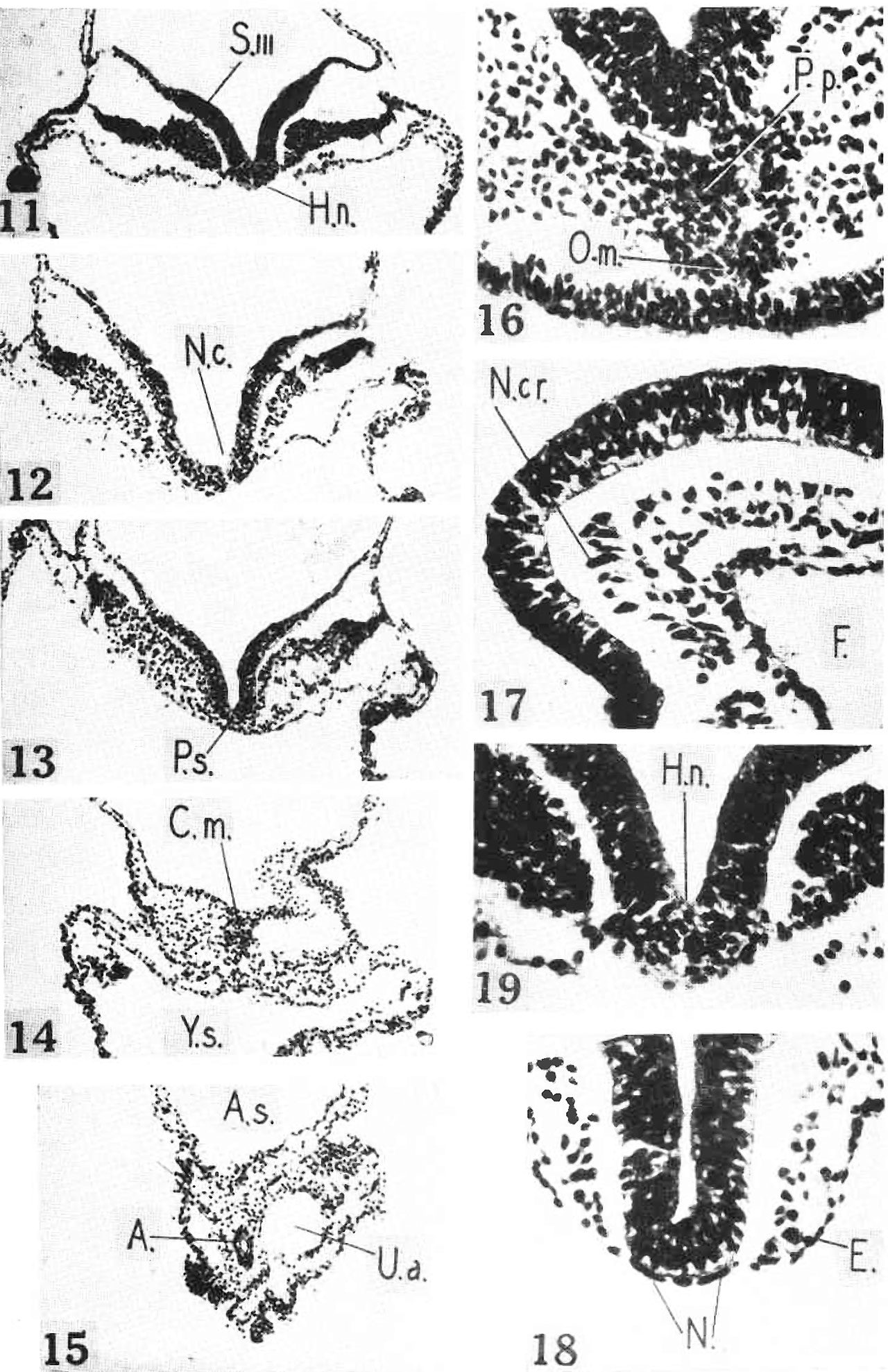
Plate 3.
Figs. 11 to 15. Photomicrographs at levels indicated in Fig. 2. Labels as in Fig. 2. x 96. Fig. 11. Through third somite and Henson's node.
Fig. 12. Through neitrenterie canal.
Fig. 13. Through middle of primitive streak.
Fig. 14. Through cloacal membrane.
Fig. 15. Through allantois and nmbilieal arteries.
Figs. 16 to 19. Photomierographs of parts of previous figtt?’6.S‘ at higher power. Labels as in Fig. 2.
Fig. 16. Portion of Fig. 3, to show prechordal plate and oral membrane. X 300. Fig. 17. Portion of Fig. 7, to show neural crest. x 300.
Fig. 18. Portion of section close to Fig. 8, to show notochord. X 300.
Fig. 19. Portion of Fig. 11, to show Henson’s node. X 300.
Reference
Shaner RF. A human embryo of two to three pairs of somites. (1945) Canad. J. Res. 23: 235-243.
Cite this page: Hill, M.A. (2024, April 28) Embryology Shaner1945 plate03.jpg. Retrieved from https://embryology.med.unsw.edu.au/embryology/index.php/File:Shaner1945_plate03.jpg
- © Dr Mark Hill 2024, UNSW Embryology ISBN: 978 0 7334 2609 4 - UNSW CRICOS Provider Code No. 00098G
File history
Click on a date/time to view the file as it appeared at that time.
| Date/Time | Thumbnail | Dimensions | User | Comment | |
|---|---|---|---|---|---|
| current | 13:14, 30 July 2017 | | 1,280 × 1,977 (475 KB) | Z8600021 (talk | contribs) | |
| 13:14, 30 July 2017 | 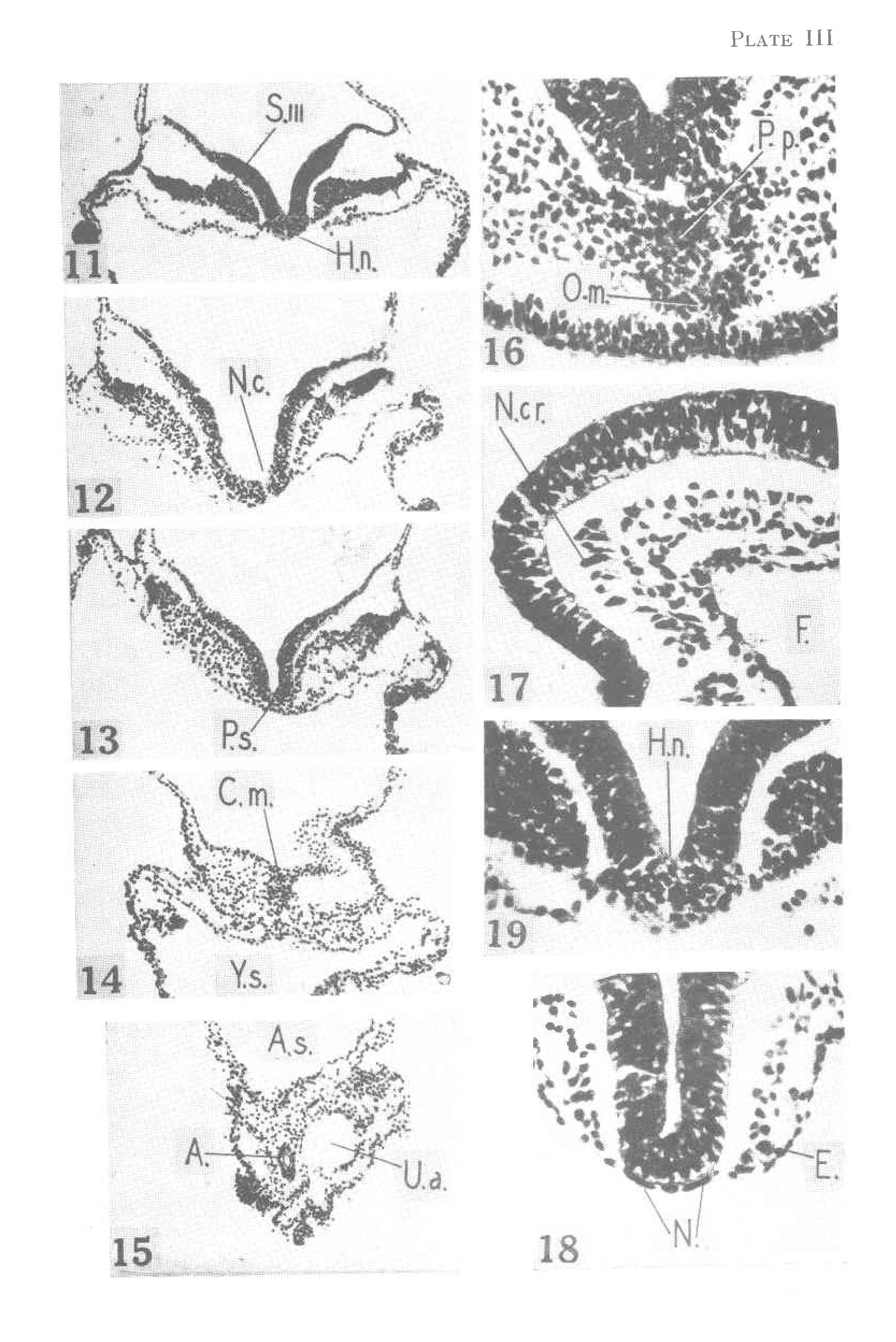 | 1,667 × 2,497 (591 KB) | Z8600021 (talk | contribs) |
You cannot overwrite this file.
File usage
The following 2 pages use this file: