File:Sgalitzer1941 fig05.jpg
From Embryology
Size of this preview: 800 × 550 pixels. Other resolution: 1,000 × 688 pixels.
Original file (1,000 × 688 pixels, file size: 104 KB, MIME type: image/jpeg)
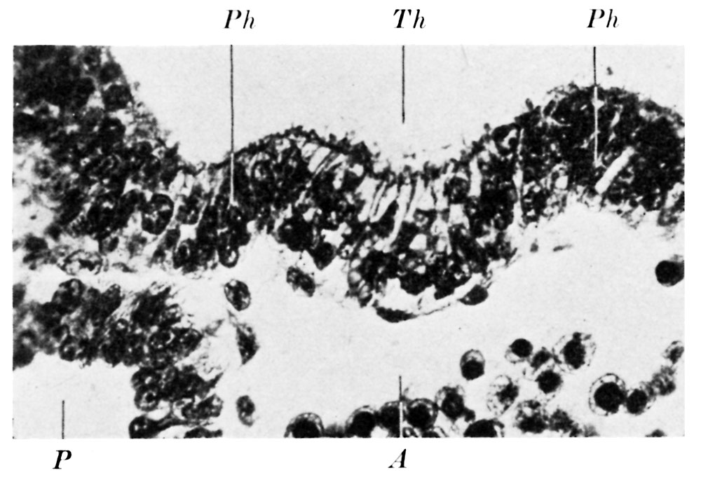
Fig. 5. Section through the thyroid anlage of embryo B
The wall of the pit represents the anlage of both the thyroid gland and the thyroglossal duct.
Reference
Sgalitzer KE. Contribution to the study of the morphogenesis of the thyroid gland. (1941) J Anat. 75(4): 389-405. PMID 17104869
Cite this page: Hill, M.A. (2024, April 26) Embryology Sgalitzer1941 fig05.jpg. Retrieved from https://embryology.med.unsw.edu.au/embryology/index.php/File:Sgalitzer1941_fig05.jpg
- © Dr Mark Hill 2024, UNSW Embryology ISBN: 978 0 7334 2609 4 - UNSW CRICOS Provider Code No. 00098G
File history
Click on a date/time to view the file as it appeared at that time.
| Date/Time | Thumbnail | Dimensions | User | Comment | |
|---|---|---|---|---|---|
| current | 10:42, 20 March 2017 | | 1,000 × 688 (104 KB) | Z8600021 (talk | contribs) | |
| 10:42, 20 March 2017 | 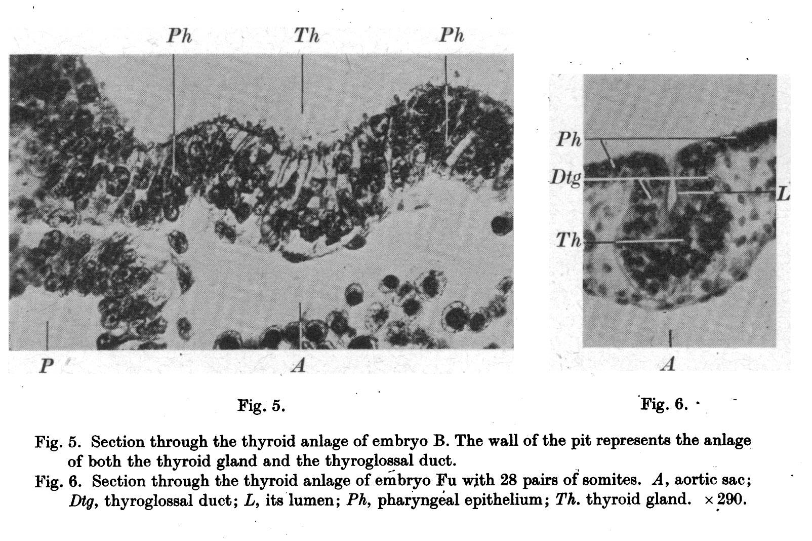 | 1,604 × 1,068 (381 KB) | Z8600021 (talk | contribs) |
You cannot overwrite this file.
File usage
The following page uses this file: