File:Sgalitzer1941 fig01.jpg
From Embryology

Size of this preview: 288 × 599 pixels. Other resolution: 577 × 1,200 pixels.
Original file (577 × 1,200 pixels, file size: 55 KB, MIME type: image/jpeg)
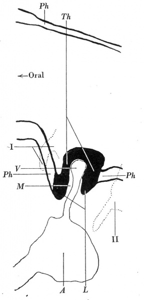
Fig 1 Mid-sagittal graphic reconstruction of the thyroid anlage of embryo F
The cavities of the first and second pharyngeal pouches of the right side have been projected on to the mid-sagital plane.
A, aortic sac; L, lipped margin of thyroid anlage; M, connective tissue core; Ph, pharyngeal epithelium; Th, thyroid anlage; F, its vessel; I, II, first and second pharyngeal pouches. x250.
Reference
Sgalitzer KE. Contribution to the study of the morphogenesis of the thyroid gland. (1941) J Anat. 75(4): 389-405. PMID 17104869
File history
Click on a date/time to view the file as it appeared at that time.
| Date/Time | Thumbnail | Dimensions | User | Comment | |
|---|---|---|---|---|---|
| current | 14:13, 19 March 2017 | | 577 × 1,200 (55 KB) | Z8600021 (talk | contribs) | |
| 14:12, 19 March 2017 | 616 × 1,728 (136 KB) | Z8600021 (talk | contribs) | ===Reference=== {{Ref-Sgalitzer1941}} |
You cannot overwrite this file.
File usage
The following 2 pages use this file: