File:Seminiferous tubule cartoon.jpg
Seminiferous_tubule_cartoon.jpg (800 × 544 pixels, file size: 92 KB, MIME type: image/jpeg)
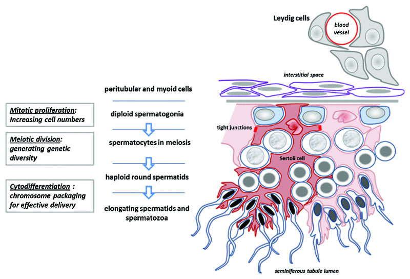
Seminiferous Tubule
Schematic diagram to illustrate the essential structure of the spermatogenic epithelium, its relation to the Leydig cells and interstitial space, and the manner in which the Sertoli cells determine the architecture of germ cell differentiation, as they progress from the tubule-enclosing basement membrane to the place of mature spermatozoa release in the tubule lumen (below).
Reference
pubmed/22553488 | PMC3341244 | Spermatogenesis
Damien Hunter, Ravinder Anand-Ivell, Sandra Danner and Richard Ivell
This is an Open Access article licensed under a Creative Commons Attribution-NonCommercial 3.0 Unported License. The article may be redistributed, reproduced and reused for non-commercial purposes, provided the original source is properly cited.
2011SPMG0032R-F2.jpg
File history
Click on a date/time to view the file as it appeared at that time.
| Date/Time | Thumbnail | Dimensions | User | Comment | |
|---|---|---|---|---|---|
| current | 08:37, 13 June 2012 | | 800 × 544 (92 KB) | Z8600021 (talk | contribs) | ==Seminiferous Tubule== Schematic diagram to illustrate the essential structure of the spermatogenic epithelium, its relation to the Leydig cells and interstitial space, and the manner in which the Sertoli cells determine the architecture of germ cell di |
You cannot overwrite this file.
File usage
The following 5 pages use this file: