File:Seminiferous tubule cartoon.jpg: Difference between revisions
No edit summary |
|||
| Line 10: | Line 10: | ||
Damien Hunter, Ravinder Anand-Ivell, Sandra Danner and Richard Ivell | Damien Hunter, Ravinder Anand-Ivell, Sandra Danner and Richard Ivell | ||
[http://creativecommons.org/licenses/by-nc/3.0/ Creative Commons Attribution-NonCommercial 3.0 Unported License]. With this license, authors retain the copyright to their work. This license also allows users to copy, distribute, transmit and adapt the work for non-commercial (educational and research) purposes. It only requires that users attribute the original authorship as well as the journal and publisher as the original source with proper citation details. Commercial rights are protected by Landes Bioscience. | |||
2011SPMG0032R-F2.jpg | 2011SPMG0032R-F2.jpg | ||
Revision as of 09:37, 16 June 2012
Seminiferous Tubule
Schematic diagram to illustrate the essential structure of the spermatogenic epithelium, its relation to the Leydig cells and interstitial space, and the manner in which the Sertoli cells determine the architecture of germ cell differentiation, as they progress from the tubule-enclosing basement membrane to the place of mature spermatozoa release in the tubule lumen (below).
Reference
<pubmed>22553488</pubmed>| PMC3341244 | Spermatogenesis
Damien Hunter, Ravinder Anand-Ivell, Sandra Danner and Richard Ivell
Creative Commons Attribution-NonCommercial 3.0 Unported License. With this license, authors retain the copyright to their work. This license also allows users to copy, distribute, transmit and adapt the work for non-commercial (educational and research) purposes. It only requires that users attribute the original authorship as well as the journal and publisher as the original source with proper citation details. Commercial rights are protected by Landes Bioscience.
2011SPMG0032R-F2.jpg
File history
Click on a date/time to view the file as it appeared at that time.
| Date/Time | Thumbnail | Dimensions | User | Comment | |
|---|---|---|---|---|---|
| current | 08:37, 13 June 2012 | 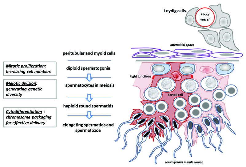 | 800 × 544 (92 KB) | Z8600021 (talk | contribs) | ==Seminiferous Tubule== Schematic diagram to illustrate the essential structure of the spermatogenic epithelium, its relation to the Leydig cells and interstitial space, and the manner in which the Sertoli cells determine the architecture of germ cell di |
You cannot overwrite this file.
File usage
The following 5 pages use this file: