File:Schaeffer1912 fig30-31.jpg: Difference between revisions
From Embryology
(===Reference=== {{Ref-Schaeffer1912}}) |
mNo edit summary |
||
| Line 1: | Line 1: | ||
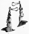
==Figs. 30 and 31 Reconstruction of the Adult nasolacrimal passages== | |||
Reconstruction of the nasolacrimal passages of an adult aged sixty-five years. X 3.2. | |||
Fig. 30 represents a medial view. | |||
Fig. 31 a lateral view of the reconstruction. Especially note the irregularity, clue to divorticula, of the nasclacrimal duct. The portions illdicated in black at the inferior extremity of the nasolacrimal duct is a portion of the inferior nasal meatus. | |||
===Reference=== | ===Reference=== | ||
{{Ref-Schaeffer1912}} | {{Ref-Schaeffer1912}} | ||
Revision as of 17:20, 18 April 2018
Figs. 30 and 31 Reconstruction of the Adult nasolacrimal passages
Reconstruction of the nasolacrimal passages of an adult aged sixty-five years. X 3.2.
Fig. 30 represents a medial view.
Fig. 31 a lateral view of the reconstruction. Especially note the irregularity, clue to divorticula, of the nasclacrimal duct. The portions illdicated in black at the inferior extremity of the nasolacrimal duct is a portion of the inferior nasal meatus.
Reference
Schaeffer JP. The genesis and development of the nasolacrimal passages in man. (1912) Amer. J Anat. 13(1): 1-18.
File history
Click on a date/time to view the file as it appeared at that time.
| Date/Time | Thumbnail | Dimensions | User | Comment | |
|---|---|---|---|---|---|
| current | 17:20, 18 April 2018 |  | 1,280 × 1,533 (86 KB) | Z8600021 (talk | contribs) | |
| 17:17, 18 April 2018 |  | 1,811 × 2,683 (213 KB) | Z8600021 (talk | contribs) | ===Reference=== {{Ref-Schaeffer1912}} |
You cannot overwrite this file.
File usage
The following page uses this file: