File:Scans of Supravalvular Aortic Stenosis and Pulmonary Stenosis.jpg
Scans_of_Supravalvular_Aortic_Stenosis_and_Pulmonary_Stenosis.jpg (665 × 436 pixels, file size: 114 KB, MIME type: image/jpeg)
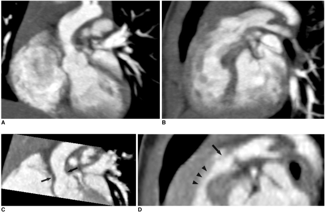
Scans of Supravalvular Aortic Stenosis and Pulmonary Stenosis in Williams syndrome Patient
--Mark Hill 15:11, 20 September 2011 (EST)Please avoid acronyms in figure titles Williams syndrome not WS. You may need to explain how this relates to the syndrome here, rather than just pasting the original figure legend.
This is a scan shows the two most common heart conditions related with Williams syndrome, Supravalvular Aortic stenosis and pulmonary stenoses in a 9 month old subject
Original legend: Combo CT scan comprised of non-ECG-synchronized spiral scan with usual scan range (A, B) prospective ECG-triggered sequential scan with narrow scan range confined to conotruncal area of heart (C, D) in 9-months-old boy with Williams syndrome. Supravalvular aortic stenosis (arrows on C) and combined valvar (arrow on D) and subvalvar (arrowheads on D) pulmonary stenoses are clearly shown on prospective ECG-triggered sequential CT images (C, D).
Dose estimates are 1.6 mSv for non-ECG-synchronized spiral scan and 0.2 mSv for prospective ECG-triggered sequential scan.
original file name: Fig. 9 Kjr-11-4-g009.jpg
Reference
<pubmed>20046490</pubmed>| PMC2799649
Copyright © 2010 The Korean Society of Radiology. This is an Open Access article distributed under the terms of the Creative Commons Attribution Non-Commercial License (http://creativecommons.org/licenses/by-nc/3.0) which permits unrestricted non-commercial use, distribution, and reproduction in any medium, provided the original work is properly cited.
- Note - This image was originally uploaded as part of a student project and may contain inaccuracies in either description or acknowledgements. Students have been advised in writing concerning the reuse of content and may accidentally have misunderstood the original terms of use. If image reuse on this non-commercial educational site infringes your existing copyright, please contact the site editor for immediate removal.
Cite this page: Hill, M.A. (2024, May 18) Embryology Scans of Supravalvular Aortic Stenosis and Pulmonary Stenosis.jpg. Retrieved from https://embryology.med.unsw.edu.au/embryology/index.php/File:Scans_of_Supravalvular_Aortic_Stenosis_and_Pulmonary_Stenosis.jpg
- © Dr Mark Hill 2024, UNSW Embryology ISBN: 978 0 7334 2609 4 - UNSW CRICOS Provider Code No. 00098G
File history
Click on a date/time to view the file as it appeared at that time.
| Date/Time | Thumbnail | Dimensions | User | Comment | |
|---|---|---|---|---|---|
| current | 14:52, 17 September 2011 |  | 665 × 436 (114 KB) | Z3331556 (talk | contribs) | original file name: Kjr-11-4-g009.jpg http://www.ncbi.nlm.nih.gov/pmc/articles/PMC2799649/ Fig. 9 Combo CT scan comprised of non-ECG-synchronized spiral scan with usual scan range (A, B) and prospective ECG-triggered sequential scan with narrow scan ran |
You cannot overwrite this file.
File usage
The following 2 pages use this file:
