File:Sabin1901 plate03.jpg

Original file (1,306 × 2,000 pixels, file size: 806 KB, MIME type: image/jpeg)
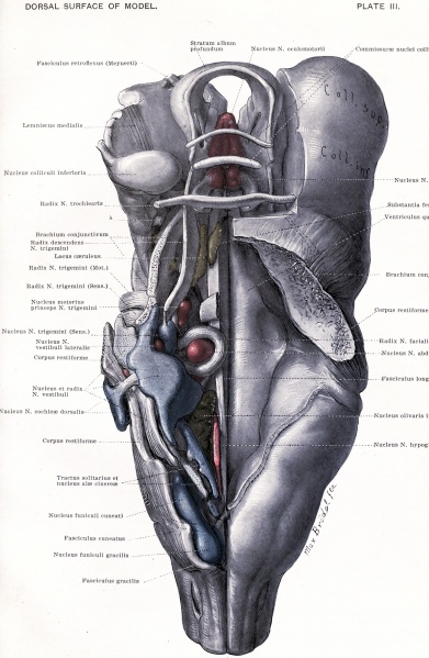
PLATE III. View of the model from the dorsal surface
On the right side is shown the floor of the fourth ventricle; on the left, the structures beneath are exposed. The position of these structures can be related to the dorsal funiculi of the spinal cord, the fourth ventricle, and the inferior and superior colliculi.
Nu. et Radix N. vestibuli: To be distinguished by the colors. The ascending root is marked by the most proximal of the three lines on the figure; the descending by the most distal line, while the nucleus N. vestibuli medialis is indicated by the middle of the three lines. The nucleus N. vestibuli superior is continuous with the medial nucleus and lies opposite the ascending root. The nucleus 1ST. vestibuli lateralis consists of two parts, one between the corpus restiforme and the ascending root, the other in the notch between the medial and superior nuclei.
Nucleus N. cochlew dor sails: The more proximal of the two lines points to the striae acusticae.
Traotus solitarius et Nu. alas cinerce: The former is in black and white, the latter in blue.
| Historic Disclaimer - information about historic embryology pages |
|---|
| Pages where the terms "Historic" (textbooks, papers, people, recommendations) appear on this site, and sections within pages where this disclaimer appears, indicate that the content and scientific understanding are specific to the time of publication. This means that while some scientific descriptions are still accurate, the terminology and interpretation of the developmental mechanisms reflect the understanding at the time of original publication and those of the preceding periods, these terms, interpretations and recommendations may not reflect our current scientific understanding. (More? Embryology History | Historic Embryology Papers) |
- Figures: Plate 1 | Plate 2 | Plate 3 | Plate 4 | Plate 5 | Plate 6 | Plate 7 | Plate 8 | Fig. 52. Key to Planes of Sections
Reference
Sabin FR. and Knower H. An atlas of the medulla and midbrain, a laboratory manual (1901) Baltimore: Friedenwald.
Cite this page: Hill, M.A. (2024, April 27) Embryology Sabin1901 plate03.jpg. Retrieved from https://embryology.med.unsw.edu.au/embryology/index.php/File:Sabin1901_plate03.jpg
- © Dr Mark Hill 2024, UNSW Embryology ISBN: 978 0 7334 2609 4 - UNSW CRICOS Provider Code No. 00098G
File history
Click on a date/time to view the file as it appeared at that time.
| Date/Time | Thumbnail | Dimensions | User | Comment | |
|---|---|---|---|---|---|
| current | 16:10, 9 June 2015 |  | 1,306 × 2,000 (806 KB) | Z8600021 (talk | contribs) |
You cannot overwrite this file.
File usage
The following page uses this file:
