File:Rugh 169.jpg

Original file (856 × 1,000 pixels, file size: 145 KB, MIME type: image/jpeg)
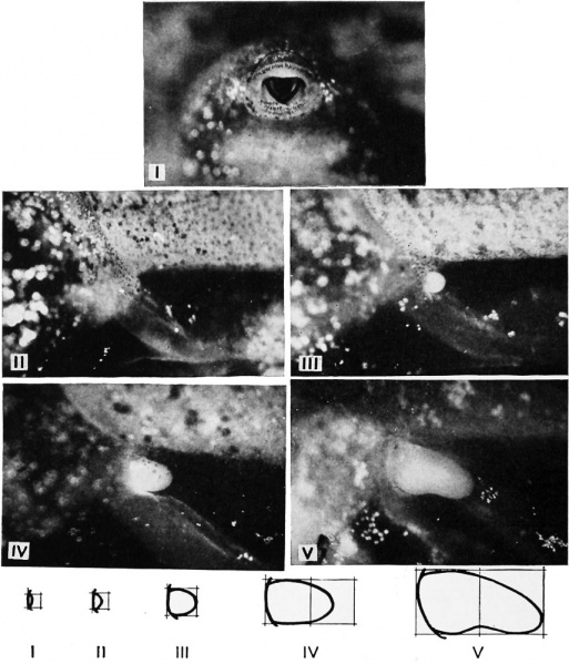
Stages in the metamorphosis of Rana pipiens
(Stage I) The oral sucker elevations have completely disappeared. Four rows of labial teeth are present, one pre-oral and three post-oral. Chromatophores have appeared and become numerous on the dorsal and lateral surfaces, and extend progressively ventrad. The limb bud is visible as a faintly circumscribed elevation in the groove between the base of the tail and the belly wall. The height of the elevation is less than half the diameter of the disk.
{Stage II) The height of the limb bud elevation (i.e., the length of the bud) is equal to half of its diameter. The first row of post-oral labial teeth is usually at the middle to form a pair of crescents. On the dorsal surface of the head the lateral line system is becoming conspicuous as pigment-free lines, especially in darkly pigmented individuals. The melanophore patches covering the gill region on either side usually meet in a narrow band ventral to the heart.
(Stage III) The length of the limb bud is equal to its diameter, and it continues to grow both in length and diameter at an approximately equal rate. This stage is relatively long in duration. (Stage IV) The length of the limb bud is equal to one and a half times its diameter.
(Stage V) The length of the limb bud is twice its diameter. The distal half of the bud is bent ventrad. There is no flattening of the tip. (Continued on facing page.)
| Historic Disclaimer - information about historic embryology pages |
|---|
| Pages where the terms "Historic" (textbooks, papers, people, recommendations) appear on this site, and sections within pages where this disclaimer appears, indicate that the content and scientific understanding are specific to the time of publication. This means that while some scientific descriptions are still accurate, the terminology and interpretation of the developmental mechanisms reflect the understanding at the time of original publication and those of the preceding periods, these terms, interpretations and recommendations may not reflect our current scientific understanding. (More? Embryology History | Historic Embryology Papers) |
Reference
Rugh R. Book - The Frog Its Reproduction and Development. (1951) The Blakiston Company.
Cite this page: Hill, M.A. (2024, April 27) Embryology Rugh 169.jpg. Retrieved from https://embryology.med.unsw.edu.au/embryology/index.php/File:Rugh_169.jpg
- © Dr Mark Hill 2024, UNSW Embryology ISBN: 978 0 7334 2609 4 - UNSW CRICOS Provider Code No. 00098G
File history
Click on a date/time to view the file as it appeared at that time.
| Date/Time | Thumbnail | Dimensions | User | Comment | |
|---|---|---|---|---|---|
| current | 18:35, 26 April 2013 |  | 856 × 1,000 (145 KB) | Z8600021 (talk | contribs) | {{Rugh1951 footer}} |
You cannot overwrite this file.
File usage
The following 2 pages use this file:
