File:Rugh 162.jpg

Original file (759 × 800 pixels, file size: 75 KB, MIME type: image/jpeg)
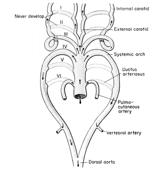
Fate of the aortic arches of the frog embryo
The aortic arches which pass through the second branchial (fourth visceral) arch become greatly enlarged and retain their connection with the dorsal aortae on both sides, to become the main systemic trunk. That portion of the dorsal aorta anterior to the second branchial arch then degenerates, so that all of the arterial blood of the first branchial arch must pass anteriorly and all of the blood of the second branchial arch must pass posteriorly. The systemic trunk on each side gives rise to the laryngeal artery ( to the larynx and muscles of the hyoid apparatus) and the oesophageal arteries (to the shoulder and brachial arteries of the limb). The paired systemic arteries join within the body to form the single dorsal aorta, sometimes called the descending aorta, which in turn gives rise to many branches. These include the renals to the glomi of the pronephros and glomeruli of the mesonephros; the large coeliac artery to the stomach, liver, pancreas, and intestine; the gonadals; the lumbars; and finally the iliacs to the hind legs.
The aortic arch which develops into the third branchial arch (fifth visceral) disappears. Those in the fourth branchial arches, however (sixth visceral), develop connections with blood islands in the skin and the lungs to become the pulmo-cutaneous arteries. In the frog, the skin represents about 60 per cent of the necessary respiratory surface. Some time after metamorphosis the connection of this fourth pair of branchial aortic arches severs connections with the dorsal aorta (now known as the systemic trunk), leaving only a vestigial strand of tissue known as the ductus Botalli or ductus arteriosus. In this manner the systemic arteries to the viscera and hmbs are separated from the respiratory arteries to the skin and lungs.
- Links: The Circulatory System
| Historic Disclaimer - information about historic embryology pages |
|---|
| Pages where the terms "Historic" (textbooks, papers, people, recommendations) appear on this site, and sections within pages where this disclaimer appears, indicate that the content and scientific understanding are specific to the time of publication. This means that while some scientific descriptions are still accurate, the terminology and interpretation of the developmental mechanisms reflect the understanding at the time of original publication and those of the preceding periods, these terms, interpretations and recommendations may not reflect our current scientific understanding. (More? Embryology History | Historic Embryology Papers) |
Reference
Rugh R. Book - The Frog Its Reproduction and Development. (1951) The Blakiston Company.
Cite this page: Hill, M.A. (2024, April 27) Embryology Rugh 162.jpg. Retrieved from https://embryology.med.unsw.edu.au/embryology/index.php/File:Rugh_162.jpg
- © Dr Mark Hill 2024, UNSW Embryology ISBN: 978 0 7334 2609 4 - UNSW CRICOS Provider Code No. 00098G
Frogitsreproduct00rugh_0253.jpg
File history
Click on a date/time to view the file as it appeared at that time.
| Date/Time | Thumbnail | Dimensions | User | Comment | |
|---|---|---|---|---|---|
| current | 17:12, 26 April 2013 |  | 759 × 800 (75 KB) | Z8600021 (talk | contribs) | {{Rugh1951 footer}} Category:Cardiovascular Frogitsreproduct00rugh_0253.jpg |
You cannot overwrite this file.
File usage
The following 2 pages use this file:
