File:Rugh 107.jpg

Original file (1,073 × 800 pixels, file size: 188 KB, MIME type: image/jpeg)
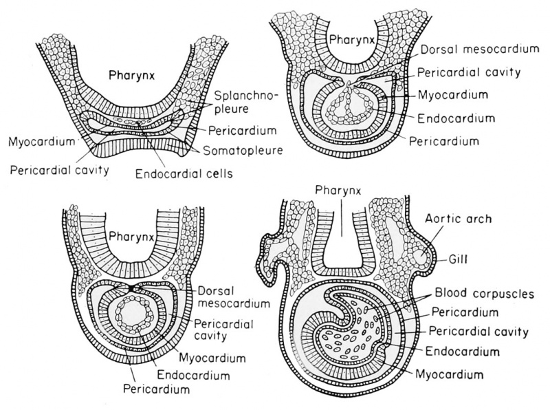
Development of the Heart of the Frog Embryo
The lateral plate mesoderm extends into the head, ventral to the pharynx, as mesenchyme. This mesenchyme becomes organized into sheets, coextensive with the more posterior lateral plate sheets of somatic and splanchnic mesoderm. These will give rise to parts of the heart. As the coelomic split occurs at the body level, there is an extension of this split into the forming heart mesoderm, which will give rise to the pericardial cavity.
The outer layer of mesoderm, corresponding to the body somatic layer, will become the pericardial membrane.
The inner layer of mesoderm, corresponding to the body splanchnic layer, will become the myocardium or heart muscle. As in the body region, the mesoderm from the two sides grows together ventrally to fuse below the foregut. Both the coelom and the continuous and related pericardial cavity arise as bilateral cavities only to fuse and form single cavities around particular organs.
The endocardium or lining of the heart arises from scattered cells of mesodermal origin found beneath the pharynx. These cells become organized into a sheet of epithelium as they are enclosed by the bilateral folds of myocardium as they come together.
| Historic Disclaimer - information about historic embryology pages |
|---|
| Pages where the terms "Historic" (textbooks, papers, people, recommendations) appear on this site, and sections within pages where this disclaimer appears, indicate that the content and scientific understanding are specific to the time of publication. This means that while some scientific descriptions are still accurate, the terminology and interpretation of the developmental mechanisms reflect the understanding at the time of original publication and those of the preceding periods, these terms, interpretations and recommendations may not reflect our current scientific understanding. (More? Embryology History | Historic Embryology Papers) |
Reference
Rugh R. Book - The Frog Its Reproduction and Development. (1951) The Blakiston Company.
Cite this page: Hill, M.A. (2024, April 27) Embryology Rugh 107.jpg. Retrieved from https://embryology.med.unsw.edu.au/embryology/index.php/File:Rugh_107.jpg
- © Dr Mark Hill 2024, UNSW Embryology ISBN: 978 0 7334 2609 4 - UNSW CRICOS Provider Code No. 00098G
File history
Click on a date/time to view the file as it appeared at that time.
| Date/Time | Thumbnail | Dimensions | User | Comment | |
|---|---|---|---|---|---|
| current | 13:39, 14 April 2013 |  | 1,073 × 800 (188 KB) | Z8600021 (talk | contribs) | {{Rugh1951 footer}} |
You cannot overwrite this file.
File usage
The following 2 pages use this file:
