File:Rugh 091.jpg

Original file (1,000 × 584 pixels, file size: 110 KB, MIME type: image/jpeg)
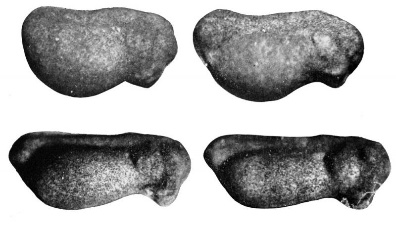
Formation of the Tail Bud
The tail bud is formed by a backward growth of tissue dorsal to the closed blastopore. As it is elongated it is provided with both a dorsal and a ventral fin, the dorsal fin being developed initially by the posterior growth of myotomes, with accompanying blood vessels and nerves, to form the tail bud.
| Historic Disclaimer - information about historic embryology pages |
|---|
 |
Reference
Rugh R. Book - The Frog Its Reproduction and Development. (1951) The Blakiston Company.
Cite this page: Hill, M.A. (2024, April 28) Embryology Rugh 091.jpg. Retrieved from https://embryology.med.unsw.edu.au/embryology/index.php/File:Rugh_091.jpg
- © Dr Mark Hill 2024, UNSW Embryology ISBN: 978 0 7334 2609 4 - UNSW CRICOS Provider Code No. 00098G
File history
Click on a date/time to view the file as it appeared at that time.
| Date/Time | Thumbnail | Dimensions | User | Comment | |
|---|---|---|---|---|---|
| current | 18:47, 12 April 2013 | | 1,000 × 584 (110 KB) | Z8600021 (talk | contribs) | |
| 18:41, 12 April 2013 |  | 1,031 × 600 (140 KB) | Z8600021 (talk | contribs) | {{Rugh1951 footer}} |
You cannot overwrite this file.
File usage
The following 2 pages use this file: