File:Rugh 037.jpg
Original file (1,180 × 900 pixels, file size: 157 KB, MIME type: image/jpeg)
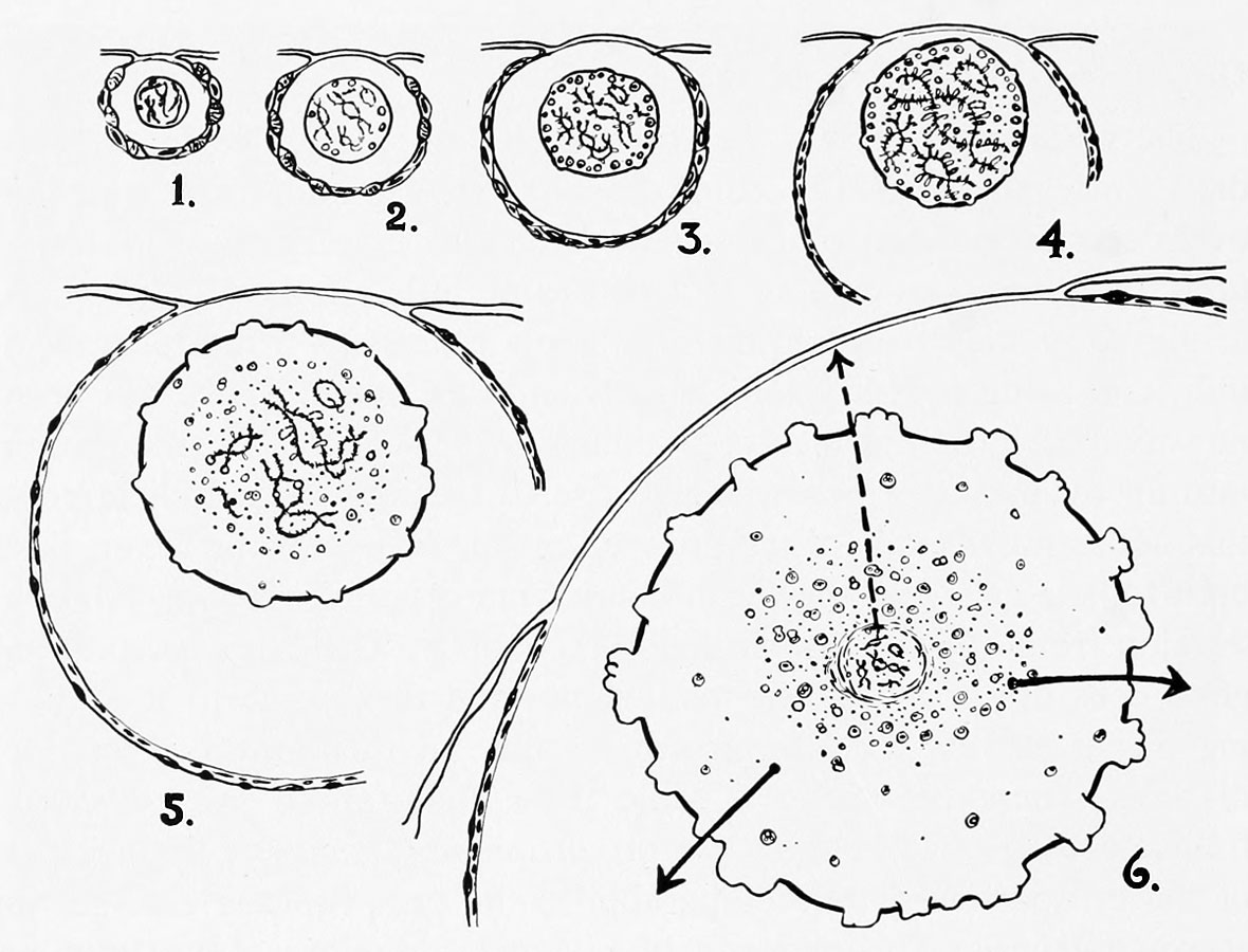
Normal nuclear growth cycle of the ovum of Rana pipiens
(Stage 1 ) Smallest follicle in which the chromosomes within the germinal vesicle can be seen.
(Stage 2) The paired chromosomes are barely visible, embedded in a nucleoplasmic gel. Egg diameters less than 200 microns.
(Stage 3) Eggs measuring from 200 to 500 microns in diameter, more detail visible through the transparent theca cells. Lateral loop production begins. Zone of large irregular nucleoli may be seen just beneath the nuclear membrane.
(Stage 4) First development of yellowbrown color and yolk. Eggs range in size from 500 to 700 microns in diameter. Chromosomes attain length of about 450 microns. For salamanders of comparable stage chromosomes measure 700 microns in length.
(Stage 5) Chromosome frame begins contraction while the nucleus continues to grow in eggs ranging in diameter from 750 to 850 microns. This is approximately half the ultimate size. Chromosomes shorten and have fewer and smaller loops. The major nucleolar production continues and sacs appear on the surface of the nuclear membrane.
(Stage 6) Egg diameter about 1.8 millimeters and germinal vesicle is of maximum size. Chromosome frame now about 1/1000 of the nuclear volume, coated by a denser substance which can be coagulated by the calcium ion. Chromosomes have shortened to 40 microns or less and have lost all large and small hyaline bodies called loop fragments.
Heavy arrows indicate the mixing of nuclear material in the cytoplasm after the breakdown of the germinal vesicle. The dotted arrow indicates migration of the central chromosomal mass toward the animal pole to become the maturation spindle for the first polar body.
Reference
From W. R. Duryee, 1950, Antu N, Y, Acad. Sci., 50, Art. 8.
| Historic Disclaimer - information about historic embryology pages |
|---|
 |
Reference
Rugh R. Book - The Frog Its Reproduction and Development. (1951) The Blakiston Company.
Cite this page: Hill, M.A. (2024, April 27) Embryology Rugh 037.jpg. Retrieved from https://embryology.med.unsw.edu.au/embryology/index.php/File:Rugh_037.jpg
- © Dr Mark Hill 2024, UNSW Embryology ISBN: 978 0 7334 2609 4 - UNSW CRICOS Provider Code No. 00098G
File history
Click on a date/time to view the file as it appeared at that time.
| Date/Time | Thumbnail | Dimensions | User | Comment | |
|---|---|---|---|---|---|
| current | 08:25, 12 April 2013 | | 1,180 × 900 (157 KB) | Z8600021 (talk | contribs) | {{Rugh1951 footer}} |
You cannot overwrite this file.
File usage
The following 2 pages use this file: