File:Retinopathy of prematurity 01.jpg
Original file (850 × 315 pixels, file size: 46 KB, MIME type: image/jpeg)
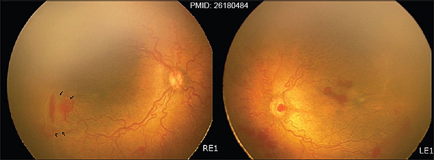
Retinopathy of Prematurity
Infant birth weight was 700 g and gestational age was 26-28 GA weeks. (RetCam images)
| Right eye | Left eye |
|---|---|
| hybrid form of retinopathy of prematurity, zone 1 disease in the right eye. Arrows show flat neovascularization |
aggressive posterior retinopathy of prematurity in the left eye. |
See also Right Eye image
Reference
<pubmed>26180484</pubmed>
Gadkari SS, Kulkarni SR, Kamdar RR, Deshpande M. Successful surgical management of retinopathy of prematurity showing rapid progression despite extensive retinal photocoagulation. Middle East Afr J Ophthalmol [serial online] 2015 [cited 2016 Feb 15];22:393-5. Available from: http://www.meajo.org/text.asp?2015/22/3/393/159778
DOI: 10.4103/0974-9233.159778
Copyright
Attribution-NonCommercial-ShareAlike 3.0 http://creativecommons.org/licenses/by-nc-sa/3.0/
Fig. 1 MiddleEastAfrJOphthalmol_2015_22_3_393_159778_u2.jpg resized, sharpened and PMID labeled.
File history
Click on a date/time to view the file as it appeared at that time.
| Date/Time | Thumbnail | Dimensions | User | Comment | |
|---|---|---|---|---|---|
| current | 15:35, 15 February 2016 | 850 × 315 (46 KB) | Z8600021 (talk | contribs) | ==Retinopathy of Prematurity== Infant birth weight was 700 g and gestational age was 26-28 {{GA}} weeks. (RetCam images) {| ! right eye ! left eye |- | hybrid form of retinopathy of prematurity, zone 1 disease in the right eye. Arrows show flat neova... |
You cannot overwrite this file.
File usage
There are no pages that use this file.