File:Ratio of alpha & beta cells at different phases of fetal development.png
Ratio_of_alpha_&_beta_cells_at_different_phases_of_fetal_development.png (537 × 600 pixels, file size: 609 KB, MIME type: image/png)
Ratio of alpha and beta cells at different phases of fetal development
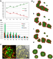
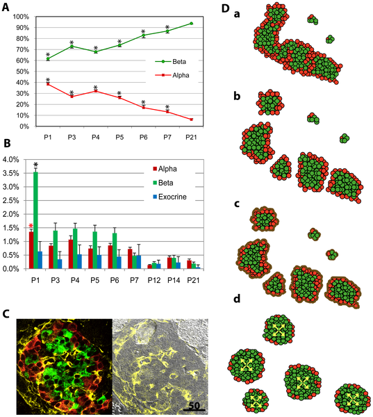
Image shows the development of the islet of langerhans and the ratio of alpha & beta cells at different phases of fetal development.
FIGURE 4: Islet formation in the neonatal pancreas. A: Increased ratio of alpha-cells to beta-cells in the neonatal pancreas. The difference was significant at all time points compared to the adult (8-mo) as a control. B: Frequency of alpha-, beta-, and exocrine-cell proliferation. Both alpha- and beta-cell proliferation at P1 was significantly increased compared to P21. C: Endocrine-cells coated with a layer of extracellular matrix (P1). Immunohistochemical staining for Insulin (green), glucagon (red) and collagen IV (yellow) is shown. Note that intra-islet blood vessels are also associated with the extracellular matrix. Scale bar is 50 µm. D: A fission model of islet formation in the neonatal pancreas. a. Endocrine cells proliferate contiguously, forming branching cord-like structures in the fetal and newborn pancreas. b. Islet formation in the neonatal pancreas may occur by fission of elongated structures composed of beta-cells and surrounding alpha-cells. The fission process appears to be random, producing islets of different size. c. Each islet is subsequently coated with a layer of extracellular matrix that stabilizes the structure. This process of islet formation may also result in small isolated clusters of pancreatic endocrine cells that persist even in the adult pancreas. d. Beta-cell mass expansion within an islet leads to an alpha-cell ratio of 5–10%.
Reference
<pubmed>19893748</pubmed>| PLos One
Copyright
© 2009 Miller et al. This is an open-access article distributed under the terms of the Creative Commons Attribution License, which permits unrestricted use, distribution, and reproduction in any medium, provided the original author and source are credited.
--Mark Hill (talk) 13:10 7 November 2014 (EST) Assessment - Figure relates to project topic pancreas development and contains reference, copyright and student template. Your legend says that it is fetal development, but all the data is mouse neonatal alpha and beta cell ration development in the pancreas. You also do not relate this back to human pancreas timeline.
- Note - This image was originally uploaded as part of an undergraduate science student project and may contain inaccuracies in either description or acknowledgements. Students have been advised in writing concerning the reuse of content and may accidentally have misunderstood the original terms of use. If image reuse on this non-commercial educational site infringes your existing copyright, please contact the site editor for immediate removal.
--Mark Hill (talk) 12:34, 11 October 2014 (EST) Please avoid using an ampersand in the file names. I have also added additional categories to your uploaded image, it is not part of the final assessment.
File history
Click on a date/time to view the file as it appeared at that time.
| Date/Time | Thumbnail | Dimensions | User | Comment | |
|---|---|---|---|---|---|
| current | 22:39, 8 October 2014 |  | 537 × 600 (609 KB) | Z3418837 (talk | contribs) |
You cannot overwrite this file.
File usage
The following 2 pages use this file:
