File:Rat kidney HIM 01.jpg

Original file (1,382 × 1,313 pixels, file size: 591 KB, MIME type: image/jpeg)
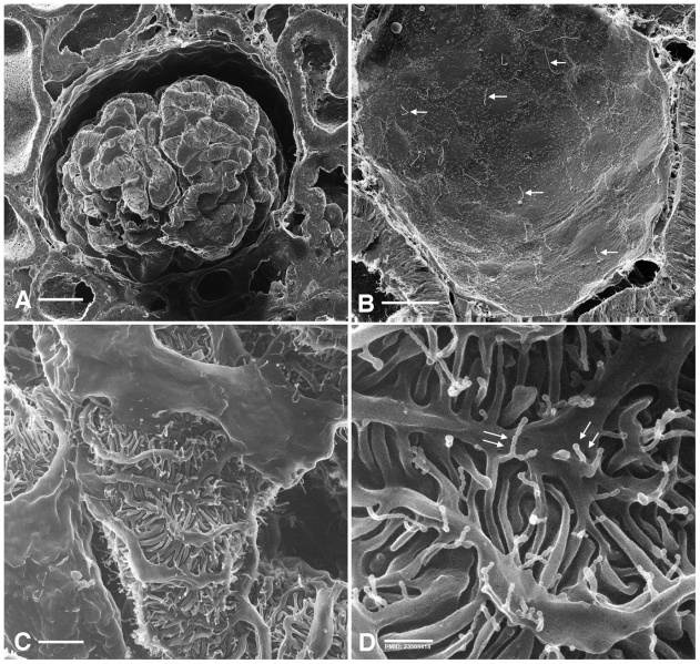
Adult Rat Kidney HIM Images
A new technique called Helium Ion Microscopy (HIM) gives similar high resolution 3D images, using a scanning beam of He+ ions, and fixed tissue without the coating required for SEM.
Reference
<pubmed>23505418</pubmed>| PMC3591388 | PLoS One.
Copyright
© 2013 Rice et al. This is an open-access article distributed under the terms of the Creative Commons Attribution License, which permits unrestricted use, distribution, and reproduction in any medium, provided the original author and source are credited.
Journal.pone.0057051.g001 Original figure 1 image adjusted in size and labelling.
File history
Click on a date/time to view the file as it appeared at that time.
| Date/Time | Thumbnail | Dimensions | User | Comment | |
|---|---|---|---|---|---|
| current | 12:51, 14 May 2014 | | 1,382 × 1,313 (591 KB) | Z8600021 (talk | contribs) | ==Adult Rat Kidney HIM Images== A new technique called Helium Ion Microscopy (HIM) gives similar high resolution 3D images, using a scanning beam of He<sup>+</sup> ions, and fixed tissue without the coating required for SEM. ===Reference=== <pubmed>... |
You cannot overwrite this file.
File usage
The following page uses this file: