File:Radford1908 fig03.jpg
From Embryology
No higher resolution available.
Radford1908_fig03.jpg (475 × 504 pixels, file size: 46 KB, MIME type: image/jpeg)
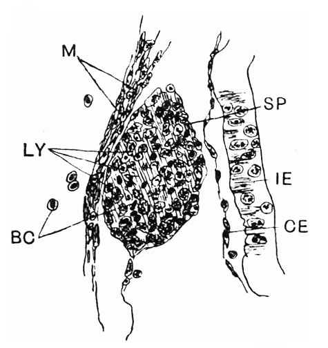
Fig. 3. Spleen in embryo of 18 mm total length
Lying between wall of mesenteric artery and wall of intestine. High power. Lymphoid cells of the spleen in various stages of division.
BC, blood-corpuscles; CE, coelomic epithelium; IE, intestinal epithelium; LY, lymphoid cells; M, primitive muscle fibres; SP, spleen.
Reference
Radford M. Development of the spleen. (1908) J Anat Physiol. 42: 288-301.
Cite this page: Hill, M.A. (2024, April 28) Embryology Radford1908 fig03.jpg. Retrieved from https://embryology.med.unsw.edu.au/embryology/index.php/File:Radford1908_fig03.jpg
- © Dr Mark Hill 2024, UNSW Embryology ISBN: 978 0 7334 2609 4 - UNSW CRICOS Provider Code No. 00098G
File history
Click on a date/time to view the file as it appeared at that time.
| Date/Time | Thumbnail | Dimensions | User | Comment | |
|---|---|---|---|---|---|
| current | 12:54, 19 July 2019 | | 475 × 504 (46 KB) | Z8600021 (talk | contribs) | |
| 12:53, 19 July 2019 |  | 735 × 844 (104 KB) | Z8600021 (talk | contribs) |
You cannot overwrite this file.
File usage
The following page uses this file: