File:Radford1908 fig02.jpg
Original file (1,031 × 1,024 pixels, file size: 168 KB, MIME type: image/jpeg)
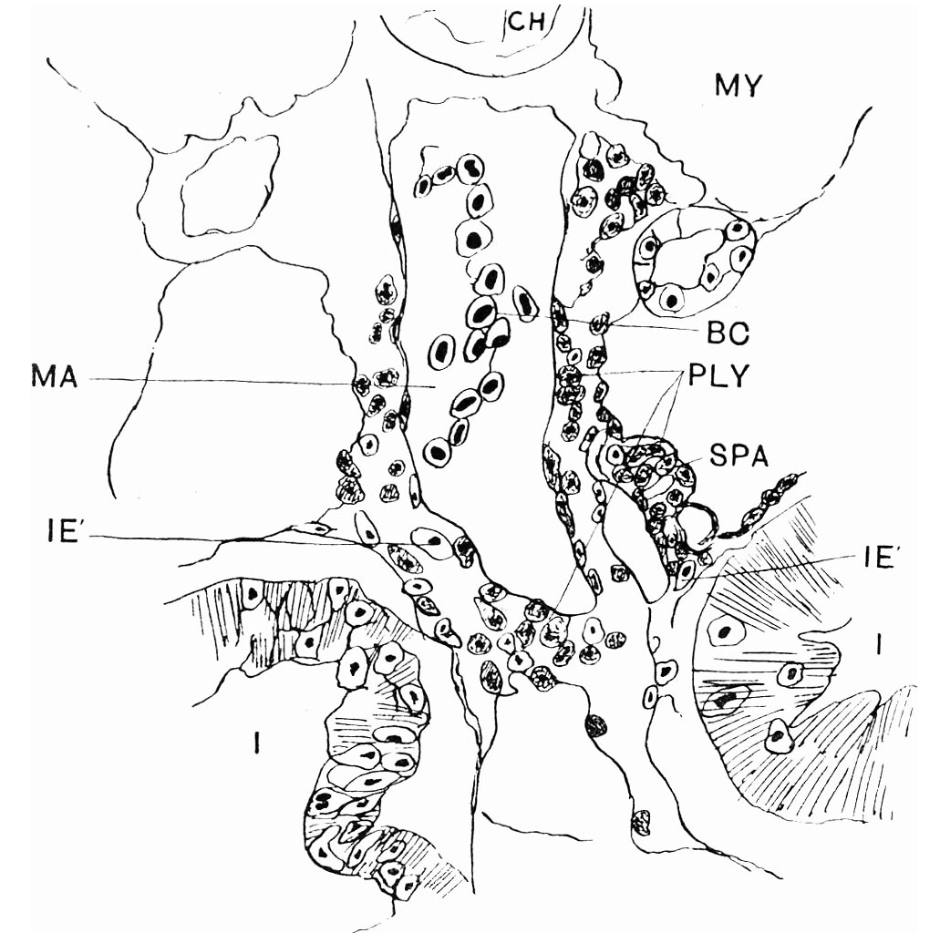
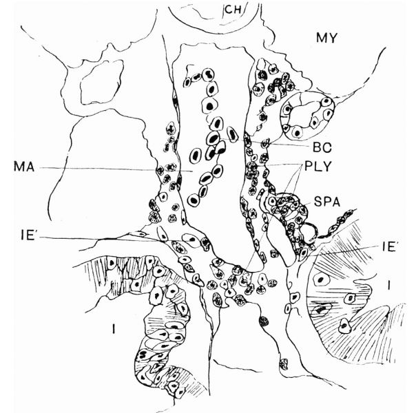
Fig. 2. Transverse section of embryo of 10 mm total length
Showing mesenteric artery with surrounding coat of lymphoid cells and the first indication of the spleen anlage. High power.
BC, blood-corpuscles; CH, notochord ; I, intestine ; IE’, strayed endodermal cells; MA, mesenteric artery; MY, myot--me; PLY, primitive lymphoid cells; SPA, spleen anlage.
Reference
Radford M. Development of the spleen. (1908) J Anat Physiol. 42: 288-301.
Cite this page: Hill, M.A. (2024, April 28) Embryology Radford1908 fig02.jpg. Retrieved from https://embryology.med.unsw.edu.au/embryology/index.php/File:Radford1908_fig02.jpg
- © Dr Mark Hill 2024, UNSW Embryology ISBN: 978 0 7334 2609 4 - UNSW CRICOS Provider Code No. 00098G
File history
Click on a date/time to view the file as it appeared at that time.
| Date/Time | Thumbnail | Dimensions | User | Comment | |
|---|---|---|---|---|---|
| current | 12:50, 19 July 2019 | | 1,031 × 1,024 (168 KB) | Z8600021 (talk | contribs) | |
| 12:47, 19 July 2019 | 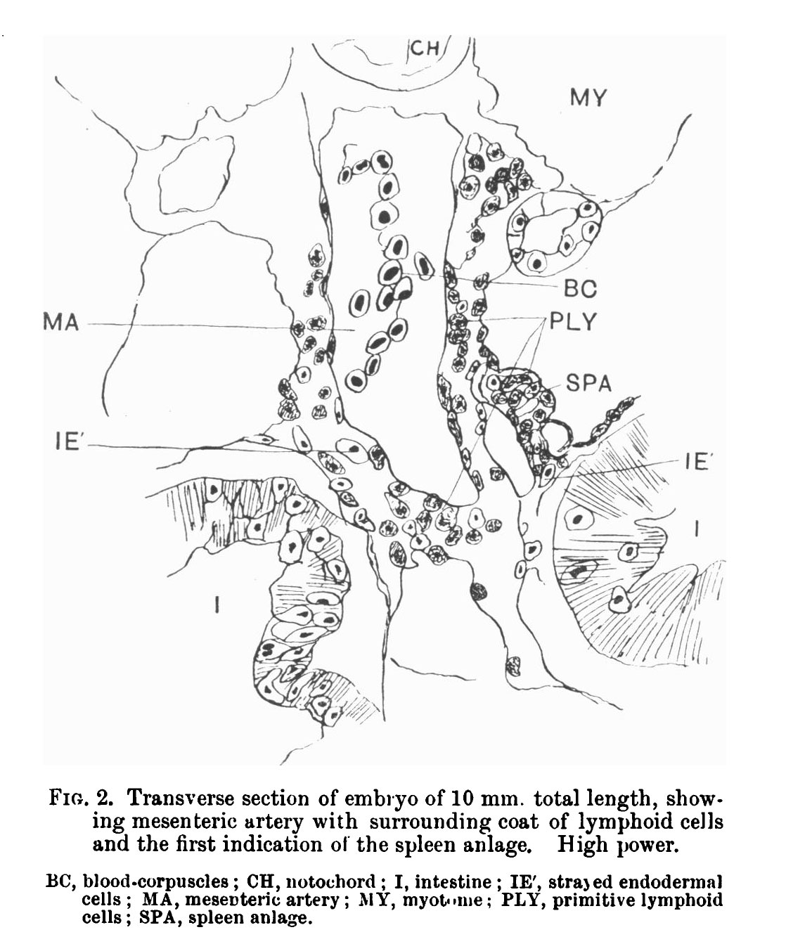 | 1,118 × 1,343 (210 KB) | Z8600021 (talk | contribs) |
You cannot overwrite this file.
File usage
The following page uses this file: