File:Quain597.jpg

Original file (811 × 1,200 pixels, file size: 204 KB, MIME type: image/jpeg)
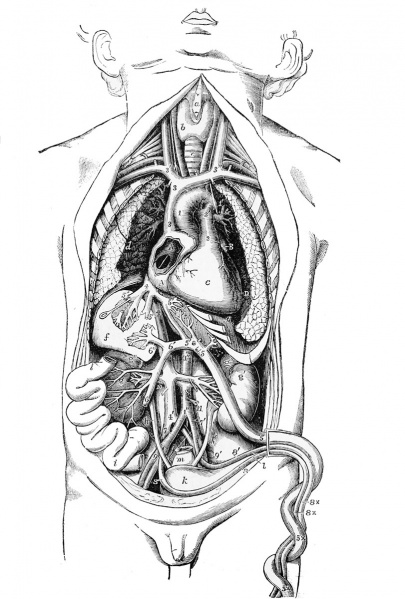
Fig. 597. Semi-diagrammatic view of the Organs of Circulation in the Foetus from Before
(from Luschka, modified, and from Nature).
a, front of the thyroid cartilage ; b, right side of the thyroid body ; c, trachea ; d, surface of the right lung turned outwards from the heart ; e, diaphragm below the apex of the heart ; /, right lobe of the liver, dissected to show ramifications of the portal and hepatic veins ; /', the middle part and left lobe of the liver in the same manner, .showing branches of the umbilical veins and ductus venosus ; g, right, fj', left kidney ; ij" , suin-arenal bodies ; h, right, li, left ureter ; i, portion of the small intestine tunied towards the side, to show the veins from it going to the portal vein ; Ic, urinary bladder ; I, is placed below the umbilicus, which is turned towards the left of the fcetus, and points by a line to the urachus ; m, rectum, divided and tied at its upjier part.
A, A. right auricle of the heart opened to show the foramen ovale : a probe, introduced through the large divided right hepatic vein and vena cava inferior, is seen passing through the fossa ovalis into the left auricle : at the lower part of the fossa ovalis is seen the Eustachian valve, to the right and inferioi'ly the auriculo-ventricular orifice ; B, the left auricular appendix ; C, the surface of the right ventricle ; D, placed on the inner surface of the left lung, points to the left ventricle.
1, ascending part of the arch of the aorta ; 1', back part beyond the ductus arteriosus ; 1", abdominal aorta; 2, stem of the pulmonary artery ; 2', the place of division into right and left pulmonary arteries and root of the ductus arteriosus : the left pueumo-gastric nerve is seen descending over the arch of the aorta ; 3, superior vena cava ; 3', right, 3", left innominate vein ; 4, stem of the inferior vena cava, between the junction of the hepatic vein and the right auricle ; 4', continuation of the vena cava inferior below ; 5, umbilical vein within the body of the foetus ; 5 x , without the bo'ly, in the umbilical cord ; 5', 5', ductus venosus ; between 5 and 5', the direct branches of the umbilical vein to the liver ; 5", 5", hepatic veins, tlu-ough one of which a probe is passed into the fossa ovalis and through the foramen ovale ; 6, vena portD3 ; 6', its left branch joining the umbilical vein; 6", its right branch; 7, placed on the right iliac vein, points to the right common iliac ai-tery; 7', left common iliac artery; 8, right, 8', left umbilical arteries coming from the internal iliac arteries ; 8 x , umbilical arteries without the body, in the umbilical cord ; 9, 9', external iliac arteries ; 10, placed below the right renal vessels ; 11, inferior mesenteric artery, above the root of which .ore seen the two spermatic arteries.
- Heart and Blood-Vessels Figures: 594. Development of Great Veins | 595. A and B. Vestige of Left Superior Cava and a Case of its Persistence | 596. Foetal Heart and Great Vessels | 597. Foetus Organs of Circulation
Reference: Quain's Elements of Anatomy. William Sharpey, Allen Thomson and Edward Albert Schafer (1878) William Wood And Co., New York.
- 1878 Elements of Anatomy: The Ovum | The Blastoderm | Fetal Membranes | Placenta | Musculoskeletal | Neural | Gastrointesinal | Respiratory | Cardiovascular | Urogenital
| Historic Disclaimer - information about historic embryology pages |
|---|
| Pages where the terms "Historic" (textbooks, papers, people, recommendations) appear on this site, and sections within pages where this disclaimer appears, indicate that the content and scientific understanding are specific to the time of publication. This means that while some scientific descriptions are still accurate, the terminology and interpretation of the developmental mechanisms reflect the understanding at the time of original publication and those of the preceding periods, these terms, interpretations and recommendations may not reflect our current scientific understanding. (More? Embryology History | Historic Embryology Papers) |
File history
Click on a date/time to view the file as it appeared at that time.
| Date/Time | Thumbnail | Dimensions | User | Comment | |
|---|---|---|---|---|---|
| current | 10:12, 8 June 2014 |  | 811 × 1,200 (204 KB) | Z8600021 (talk | contribs) | {{Quain header}} |
You cannot overwrite this file.
File usage
The following page uses this file:
