File:Quain594.jpg
Original file (592 × 1,000 pixels, file size: 87 KB, MIME type: image/jpeg)
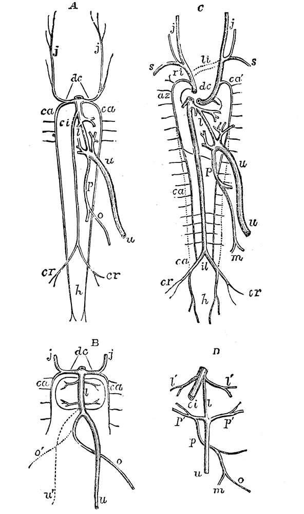
Fig. 594. Diagrams illustrating the development of the Great Veins
(after Kollliker)
A, plan of the principal veins of the fretus of about four weeks, or soon after the first formation of the vessels of the hver and the vena cava inferior.
B, veins of the liver at a somewhat earlier period.
C, principal veins of the foetus at the time of the first estabhshment of the placental circulation.
D, veins of the liver at the same period.
dc, the right and left ducts of Cuvier ; ca, the right and left cardinal veins ; j. j,ihe jugular veins ; s, the subclavian veins ; az, the azygos vein ; u, the umbilical or left umbilical vein ; u', in B, the temporary right umbilical vein ; o, the omphalo-meseuteric vein ; o', the right omphalo-mesenteric vein ; m, the mesenteric veins ; p, the portal vein ; p', p', the vente advehentes ; I, the ductus venosus ; V, I', the hepatic veins ; c'l, vena cava inferior ; il, the division of the vena cava inferior into common iliac veins ; cr, the external iliac or crural veins ; h, the hypogastric or internal iliac veins, in the line of continuation of the primitive cardinal veins.
In C, li, in dotted lines, the transverse branch of communication between the jugular veins which forms the left innominate vein ; ri, the right innominate vein ; ca, the remains of the left cardinal vein by which the superior intercostal veins fall into the left innominate vein ; above lo, the obliquely crossing vein by which the hemiazygos joins the azygos vein.
- Heart and Blood-Vessels Figures: 594. Development of Great Veins | 595. A and B. Vestige of Left Superior Cava and a Case of its Persistence | 596. Foetal Heart and Great Vessels | 597. Foetus Organs of Circulation
Reference: Quain's Elements of Anatomy. William Sharpey, Allen Thomson and Edward Albert Schafer (1878) William Wood And Co., New York.
- 1878 Elements of Anatomy: The Ovum | The Blastoderm | Fetal Membranes | Placenta | Musculoskeletal | Neural | Gastrointesinal | Respiratory | Cardiovascular | Urogenital
| Historic Disclaimer - information about historic embryology pages |
|---|
 |
File history
Click on a date/time to view the file as it appeared at that time.
| Date/Time | Thumbnail | Dimensions | User | Comment | |
|---|---|---|---|---|---|
| current | 10:50, 8 June 2014 | | 592 × 1,000 (87 KB) | Z8600021 (talk | contribs) | {{Quain figures9}} |
You cannot overwrite this file.
File usage
The following page uses this file: